sprains-and-strains

Sprains and Strains: causes, symptoms, treatment

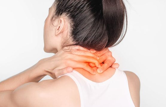
Sprains and strains are common injuries that affect muscles and ligaments. Most can be treated at home without seeing a primary care physician.

What is the difference between sprain and strain? The ligaments, muscles, or tendons that line the bones and wrists can be affected if the movements we make are not correct (sudden or violent movements of the bones).

Sprain
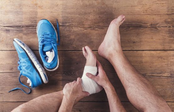
It consists in the violent stretching of the articular ligaments, but without a rupture or tearing. Sprain occurs in the ankle, knee or wrist.

We recognize a sprain after painful swelling, the movements can be done, but they are not natural and even bruises can occur.

Until he arrives at the hospital, the wounded man can be given ice packs or we can immobilize his area by wrapping it with an elastic bandage or bandage. At the hospital he will be consulted by a specialist doctor and an X-ray will be taken.

The treatment of the sprained ankle consists in putting a bandage or a splint for a period of 2 weeks or in the most painful case it is put in plaster.
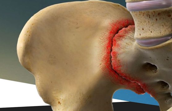
Strain
It means moving a bone in the joint. The dislocation receives the name of the region in which it occurred: shoulder, foot, elbow.

Strain occurs by falling on the shoulder, elbow, foot (by landing from the jump on the outer edge of the foot). It is more common in athletes (sports injuries) and tourists.

First aid in case of strain of the foot is done by pulling the foot as it is done when removing the boot, the knee must be in flexion to allow relaxation of the Achilles tendon. Arriving at the hospital, he was received by a medical specialist.

CONTENT:

  1. Causes
  2. Symptoms
  3. Treatment


Causes of Sprains and Strains

The movements of the ankle and knee joints, performed suddenly and forcing the physiological limits of the body, can cause a minor trauma, by stretching the ligaments that connect the bones of the foot and those of the leg. In critical cases, the inversion, the most common cause of sprain, is generated by rotating the ankle inward, simultaneously with bringing the sole of the foot inward.

An even more painful form of a sprain is caused by twisting the upper ligaments and those outside the calf. External rotation and abduction of the ankle lead to sprain by eversion, a form of trauma that may require surgery. Most often, you can have the unpleasant experience of a sprain on a walk in the mountains, due to choosing the wrong pair of shoes, or on the football field, during a man-to-man dribble.


Symptoms of Sprains and Strains

The intensity of the symptoms depends on the severity of the lesions. The first symptom of a sprain is pain, although the painful sensation may occur with some delay after the injury, because the inflammatory process takes time. It is not excluded that the moment of ligament overload is not aware.

Local pain and tenderness, but also exacerbation of painful sensations when mobilizing the joint are specific symptoms of sprains. Swelling of the affected area is sometimes associated with bleeding in the ligaments. Deep bleeding is associated with bruising of the skin, but it can take up to 24 hours for it to become apparent.

The combination of pain and swelling of the joint causes the body to protect the injured area, which leads to functional difficulties through self-preservation. Sprains located at the ankle or knee are associated with a limited ability to move the joint, its mobilization being associated with intense pain.

In some situations, all these symptoms may be associated with a numbness, and in the case of severe sprains, at the time of occurrence, it is possible to hear or feel, when mobilizing lightly, a joint click (a crack).


Treatment of Sprains and Strains

Simple forms of sprain can be treated at home. In their case, it is necessary to immobilize the injured area and avoid forcing it. It is sometimes necessary to rest for a few days and limit the usual activities, which put pressure on the affected joint.

Ice packs are useful. They are kept on the joint for about 10-15 minutes, four to eight times a day. It is also useful to massage with an ice cube on the joint, through circular movements.

Compression of the joint is useful in reducing inflammation. Elastic bandages can be used on the affected area and on the extremities to prevent distal inflammation. The compressive bandage soothes pain, but should not be excessively tight, uncomfortable or disrupt blood circulation. Also to reduce the inflammation of the limb with the affected joint, it must be kept high, at a height above the patient’s heart.

In more severe cases, in which the ligaments have been partially torn, it is necessary to immobilize the ankle with a cast of plaster or orthosis. The time required for healing is set by the orthopedist. For very severe sprains, with torn ligaments, associated with bleeding, surgery is required.

Therapy should not be limited to reducing pain, reducing edema and eliminating bruising, but should be continued after the acute phase is over, with medical gymnastics and physiotherapy, so that the affected joint is restored to normal parameters or as close as possible to the condition before injury.

The recovery protocol is necessary to reduce the sensitivity of the joint, but also in order to prevent recurrence, whose risk is relatively high, which can lead to motor deficit or a compensatory position that causes, over time, other postural disorders. It is important to know that the pain passes before the affected joint is completely restored, and medical gymnastics is essential for complete healing.

Manual therapy is recommended especially in case of severe sprains and recurrences or after periods of immobilization. It is important to say that these joint injuries, especially in the knee, are considered favorable factors for osteoarthritis.

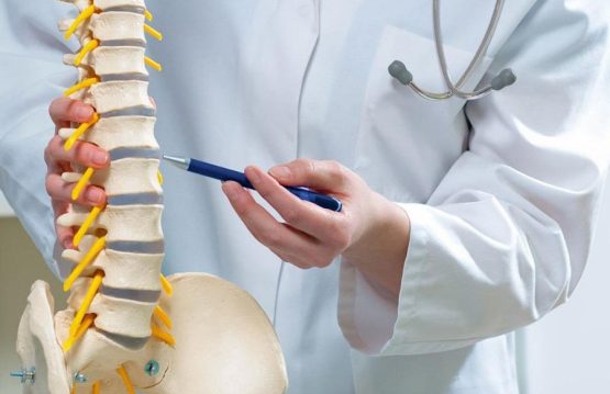
Regardless of the severity of the sprain, it is recommended to consult a specialist, in order to benefit from a correct evaluation and personalized therapeutic indications.

The duration of recovery depends on the severity of the sprain. In mild cases, it can last one to two weeks, while in more severe cases, it can reach 10-12 weeks. Surgical repairs, necessary for completely torn ligaments, have the longest recovery period and are followed by postoperative therapy.

The presentation to the doctor is absolutely necessary, if the symptoms worsen after the application of cold compresses and compression of the joint, with elastic band, the second or third day after the trauma.

spondylosis

Spondylosis: causes, symptoms, treatment

Spondylosis is also known as spinal osteoarthritis – a fairly common condition, but it is usually not serious, although it can be quite painful. As with many other terms for describing spinal problems, spondylosis is more of a descriptive term than a clinical diagnosis. Literally, it can be translated to mean both pain and degeneration of the spine, regardless of what causes the pain or where the degeneration occurs.

Spondylosis is becoming more common. Over 80% of people over the age of 40 have spondylosis in radiographic studies.

CONTENT:

  1. Causes
  2. Symptoms
  3. Treatment


Causes of Spondylosis

The bones and cartilage in the cervical spine are more prone to damage due to various causes. These include:

  • Bone spurs – are some bone growths that occur when the body develops excess bones to make the spine stronger.

However, these bones can put pressure on delicate areas of the spine, such as the spinal cord and nerves, which is why the pain occurs.

  • Dehydrated spinal discs – the bones of the spine have discs between them, which are similar to thick pillows, which absorb the shock of lifting, twisting and other activities. The gel material in these discs can dry over time. This causes the bones (vertebrae of the spine) to rub more, which can be painful.

This process can begin at the age of 30.

  • Disc herniation – the vertebral discs may develop cracks, allowing internal cushioning material to drain. It can press on the spinal cord and nerves, leading to symptoms such as numbness of the arm and pain radiating down one of the arms.
  • Injury – a trauma to the neck (due to a fall or a car accident, for example) can speed up the aging process.
  • Ligament stiffness – the hard cords that connect the bones of the spine can become even harder over time, which affects the movement of the neck.
  • Overload – some activities or hobbies involve repetitive movements or difficult efforts that can put pressure on the spine, resulting in its early wear.


Symptoms of Spondylosis

These symptoms can range from mild to severe, and can become chronic. These may include:

Neck (cervical area)

  • The pain that appears and disappears
  • Pain that spreads to the shoulders, arms, hands or fingers
  • Stiffness in the morning in the neck or shoulder, or a limited range of motion
  • Numbness of the neck or shoulder
  • Weakness or tingling in the neck, shoulders, arms, hands or fingers
  • Headache felt in the nape of the neck
  • Loss of balance
  • Difficulty swallowing (This is rare, but may occur if the spinal cord is compressed.)


Chest area

  • Bending and enlarging the body triggers pain
  • Stiffness in the back when getting out of bed in the morning


Lower back

  • Pain that appears and disappears
  • Stiffness in the back in the morning
  • Pain that decreases after rest or exercise
  • Weakness or numbness in the lower back
  • Sciatica (mild to severe pain in the foot)
  • Weakness, numbness or tingling in the back, legs or soles
  • Difficulty walking
  • Intestinal or bladder disorders (this is rare, but may occur)


Treatment of Spondylosis

Often, methods that do not involve surgery are very effective.

A physiotherapist is usually helpful for people with cervical spondylosis. Physical therapy helps to stretch the muscles of the neck and shoulders, which, in the end, will lead to pain relief.

Therapeutic massage is often indicated before physiotherapy sessions, to calm and relax the area, combat painful muscle contractions, stimulate blood circulation and muscle toning.

Your doctor may recommend medications such as anti-inflammatory drugs, narcotics, antiepileptics, steroid injections or topically applied gels.

If the patient’s condition is severe and he does not respond to other forms of treatment, surgery may be needed. This may involve removing the bone spurs, parts of the neck bones or herniated discs to give more space to the spinal cord and nerves.

Surgery is rarely necessary for cervical spondylosis. However, the doctor recommends this method if the pain is severe and if the patient’s ability to move his arms is affected.

Natural remedies
There are several natural remedies that may help those suffering from cervical spondylosis. These include:

  • Ginger tea;
  • Devil’s claw supplements;
  • White willow bark extract supplements.


Although they can help reduce pain and inflammation, they should be taken with your doctor’s consent. Even if it is an herbal tea, it can have unwanted effects if it interacts with another commonly used medicine prescribed by your doctor.

Spondylolisthesis

Spondylolisthesis: causes, symptoms, treatment

Spondylolisthesis is the anterior slip of one vertebra to the rest of the vertebrae, more precisely the vertebra changes its initial position and leaves the alignment of the other adjacent vertebrae.

Spondylolisthesis is classified according to the causes of its occurrence and is of five types: congenital, isthmic, degenerative, traumatic and pathological.


CONTENT:

  1. Causes of Spondylolisthesis
  2. Symptoms of Spondylolisthesis
  3. Treatment of Spondylolisthesis


Causes

  • congenital defect in the spine (usually occurs a few years after birth);
  • acute trauma to the back;
  • exaggerated repetitive extensions (common in certain sports);
  • muscle atrophies;
  • degenerative diseases of the spine.


Symptoms

Other people suffer from pain, especially under stress and during certain movements. The pain caused by spondylolisthesis can spread from back to front in a belt shape. There is also a feeling of instability in the spine.

Especially in the morning, when the back muscles are relaxed, the pain is strong. In severe cases, there are reflex, sensory and motor disorders, which can extend to the legs. These symptoms occur when the vertebra squeezes a nerve root through a spondylolisthesis.

However, there are no specific gliding symptoms, as the symptoms may be similar to those of other back problems, such as herniated discs.

In the congenital form of patients with spondylolisthesis, usually the symptoms are mild or mild, being a slow progressive process. So the nerves have the opportunity to adapt to changed circumstances.


Treatment

Physical and rehabilitation therapy
It should not be initiated until after a period of rest and once the pain with daily activities has diminished. Physical regimens reduce the stress by extension of the lumbar spine and promote a nonlordotic position. It consists of exercises for stretching the abdominal muscles, wearing harnesses and strengthening the lumbodorsal fascia.

Thoracolombosacral orthosis trimming provides relief for those who do not respond to rest or whose daily activities cause symptoms. This type of rod is effective for patients with less than 50% slip. The stem is worn for 3-6 months. If the slip is below 50% but the patient is symptomatic, non-interventional therapy is recommended: stretching exercises, antilordotic rod, modification of activities. If the pain continues, spinal fusion is recommended.

Surgical therapy
Surgery is indicated for immature skeletal patients with slipping over 30-50% because they are at risk of progression with neurological deficit and persistent pain. If the pain does not resolve in 6-12 weeks with rest and immobilization, surgery is recommended. Spondylolysis with low-grade spondylolisthesis can be treated noninterventionally.

Intervention options include direct repair of spondylolytic defect, fusion, reduction and fusion, and vertebrectomy. The best results are seen in patients with lithic defect. Disc degeneration is a relative contraindication. Slips over 2 mm decrease the success of surgical repair.

In situ fusion at the affected level is the standard criterion for surgical treatment for most patients in whom conservative therapy fails. In situ fusion is recommended for patients with low-grade, persistent, symptomatic spondylolisthesis and for those who are not candidates to repair the defect.

Decompression and fusion are performed in cases of hard compression in the presence of intestinal or bladder dysfunction or significant motor deficits. Decompression is never performed without concomitant fusion. Fixing the pedicles with screws allows rapid mobilization and early ambulance after decompression and fusion. Fixation can be beneficial in repairing pseudoarthrosis and preventing progressive slipping.

The reduction of spondylolisthesis is performed by the closed or open procedure. The reduction serves to correct the lumbosacral kyphosis and decrease the sagittal translation. Vertebrectomy can be used to treat spondyloptosis and as an alternative to reduction or fusion in situ. The postoperative rate of neurological deficit is 25%

spinal-tumors

Spinal Tumors: causes, symptoms, treatment

A spinal tumor is a mass of abnormal tissue that develops in the spine, either in the spinal cord (spinal cord tumors) or in the vertebrae (vertebral tumor or extradural tumor). The cells that make up the tumor are abnormal cells, which grow outside the usual mechanisms of cell growth control. Spinal tumors can cause pain, neurological problems and sometimes paralysis.

A spinal tumor can be life-threatening and can cause permanent disability. Treatment of spinal tumors may include surgery, radiation therapy, chemotherapy and other medications, most often combinations of these. Evidence indicates surgical resection of spinal cord tumors as the gold standard in the treatment of these conditions, chemotherapy and radiation being used for tumors with a high degree of infiltration into neighboring tissues or for recurrent tumors.

They can be malignant tumors (cancerous) or benign tumors (non-cancerous) that can develop in the spinal cord or surrounding areas. A malignant spinal tumor contains cancer cells, grows rapidly, invades nearby healthy tissues and can be life-threatening.

A benign tumor does not contain cancer cells, rarely invades nearby tissues or spreads to other parts of the body and usually does not grow again after it is removed. However, even if some such tumors are not cancerous, they can compress the spinal cord or nerve roots and cause symptoms. Some common benign spinal tumors are osteoma, osteoblastoma, hemangioma, neurofibroma, and osteochondroma. Fortunately, spinal tumors are relatively rare.


CONTENT:

  1. Causes of Spinal Tumors
  2. Symptoms of Spinal Tumors
  3. Treatment of Spinal Tumors


Causes

The exact cause of spinal tumors is unknown. It is suspected that certain defective genes are involved, but it is not known whether such genetic defects are inherited or simply develop over time. These could be caused by environmental factors, such as exposure to certain chemicals.

However, in some cases, spinal tumors are linked to known inherited syndromes, such as neurofibromatosis 2 and von Hippel-Lindau disease.


Symptoms

Because spinal tumors can affect the spinal cord or nerve roots, blood vessels, or vertebrae, the symptoms are different and occur especially as the tumors grow. Possible signs and symptoms include:

  • Pain at the site of the tumor due to its growth
  • Back pain, which often radiates to other parts of the body
  • Lack of sensitivity to pain, heat or cold
  • Fecal or urinary incontinence
  • The weight of walking, which sometimes leads to falls
  • Back pain that gets worse at night
  • Loss of sensitivity or muscle weakness, especially in the arms or legs
  • Muscle weakness, which can be moderate or severe, in different parts of the body.
  • Back pain is an early symptom of spinal tumors. The pain can spread to the hips, legs, soles or arms and can worsen over time – even with treatment.


Spinal tumors have varying degrees of evolution, depending on the type of tumor.

Vertebral tumors can weaken bone structure, predisposing the spine to fractures due to minor trauma.


Treatment

The goal of treatment is to reduce or prevent nerve damage caused by pressure on the spinal cord. The faster the symptoms develop, the faster treatment is needed to prevent permanent damage.

Ideally, the goal of treatment is to completely remove the tumor, but this goal can be complicated by the risk of permanent damage to the spinal cord and surrounding nerves. Physicians must also take into account age and general health. Corticosteroids (dexamethasone) can be given to reduce inflammation and swelling around the spinal cord.

Emergency surgery may be needed to improve spinal cord compression. Some tumors can be completely removed. In other cases, only part of the tumor can be removed to relieve pressure on the spinal cord. Chemotherapy has not been shown to be effective against most primary tumors, but may be recommended in some cases, depending on the type of tumor.

The type of non-surgical treatment depends on many factors, including the type of tumor (benign or malignant), the stage of the disease, the goal of treatment (eg, pain reduction, healing), the patient’s life expectancy, and general health.

Surgery
It may be recommended to remove the tumor if it is limited to only a portion of the spine. During surgery, there may be a risk of damage to nearby nerves or the spinal cord. Surgical resection can be combined with radiation or chemotherapy to remove residual cancer cells.

Surgery to treat a spinal tumor may be warranted when a specific biopsy is needed, if the tumor causes compression of the spinal cord or nerves, if the neurological deficit progresses, the pain does not respond to non-surgical treatment, we are talking about the destruction of the vertebrae or develops spinal instability.

But even with the latest technological advances in surgery, not all tumors can be completely removed. When the tumor cannot be completely removed, surgery may be followed by radiation or chemotherapy. Recovery after surgery on the spine can take weeks or even longer, depending on the procedure. There may be a temporary loss of sensation or other complications, including bleeding and nerve tissue damage.

Radiotherapy
It is done either after surgery for spinal cancer or alternatively to treat spinal cancer that is not operable.

Chemotherapy
One or more anti-cancer drugs are given to kill the tumors. Some of the commonly used drugs are methotrexate, doxorubicin, cyclophosphamide, carboplatin, ifosfamide. These drugs are given either orally, injected intravenously or intramuscularly, or injected directly into the affected area (including injection into the cerebrospinal fluid if the tumor affects the spinal cord or spinal nerves).

Palliative therapy
Pain therapy is also called palliative treatment – the goal is to relieve pain, reduce symptoms and prevent complications. These are treatments that do not cure the disease, but rather improve the patient’s quality of life. Treatment may include anti-inflammatory drugs and various forms of treatment for pain, including morphine. Physical therapy may be prescribed to help a patient regain flexibility and muscle strength. In addition, your doctor may recommend nutritional support.


spinal-stenosis

Spinal Stenosis: causes, symptoms, treatment

Spinal stenosis is a degenerative condition of the spine characterized by narrowing of the spinal canal through which the spinal cord and spinal nerves pass. Narrowing of the canal compresses the spinal nerves and spinal cord into cervical and thoracic area and only the spinal nerves in the lumbar area, because the spinal cord ends in normally at the level of the L1 vertebra. Canal stenosis can be most commonly seen in the lumbar area and cervical.

Spinal stenosis is caused by degenerative changes, wear and tear. In severe cases of stenosis, intervention surgery may be recommended.

The spinal canal is located in the spinal cord and is the one that protects the spinal cord. The wall of the spinal canal consists of several structures: the vertebral discs, the intervertebral joints, the flavum ligament, which stabilizes the spine in length.

The three main types of spinal stenosis are:
– cervical stenosis – narrowing of the spinal canal at the neck;
– lumbar stenosis – narrowing of the lumbar spinal canal, the space inside the lower part of the spine that transports the nerves to the legs;
– thoracic stenosis – this is rare and affects the middle / upper part of the spine.

CONTENT:

  1. Causes of Spinal Stenosis
  2. Symptoms of Spinal Stenosis
  3. Treatment of Spinal Stenosis


Causes

With age, natural degenerative processes occur throughout the body. Osteoarthritis is the most common cause of spinal stenosis, causing the formation of bone spurs that can grow in the spinal canal, compressing the nerves.

Other causes may include:
– disc herniations – soft discs located between the vertebrae that act as shock absorbers tend to dry out with age; cracks on the outside of a disc can allow some of the soft inner material to escape and press on the spinal cord or nerves;
– spinal injuries – car accidents and other traumas can cause dislocations or fractures of one or more vertebrae; displaced bones from a fracture of the spine can damage the contents of the spinal canal;
– tumors – abnormal growths can form inside the spinal cord, in the membranes that cover the spinal cord or in the space between the spinal cord and vertebrae; these are less common and can be identified on spinal imaging;
– Paget’s disease – a condition in which the bones grow abnormally large and fragile, the result being a narrowing of the spinal canal.


Symptoms

There may be people who do not show symptoms, the presence of spinal stenosis being highlighted on an MRI scan or a computed tomography. When they occur, they often begin gradually and worsen over time. Symptoms vary depending on the location of the stenosis and the nerves that are affected.

In the case of cervical stenosis:
– numbness or tingling in the hand, arm, foot or sole;
– weakness in the hand, arm or leg;
– problems with walking and balance;
– sore throat;
– in severe cases, loss of sphincter control and urinary incontinence.

In case of lumbar stenosis:
– numbness or tingling in the legs, back or buttocks;
– pain or cramps in one or both legs when standing for long periods of time or when moving, pain that decreases in intensity when you sit or lean forward;
– loss of control in the arms or legs which can lead to gait or holding objects;
– back pain.


Treatment

Therapy for spinal stenosis begins with conservative measures, similar to disc herniation: medical gymnastics, anti-inflammatory drugs, possibly cortisone injections into the spinal canal. In addition, you can opt for certain corsets that support maintaining the spine in a certain position to avoid applying pressure on the spinal canal and nerves.

If these measures do not significantly improve the symptoms of spinal stenosis, surgery is required. Depending on the severity of the symptoms, minimally invasive interventions and classic operations are possible. In the case of minimally invasive interventions, following spinal anesthesia, spacer devices are installed that extend the spinal canal and stretch the spine. Given that this procedure is relatively new, long-term results are not yet available.

Surgery: good long-term results

Usually, decompression of nerve roots is necessary to treat spinal stenosis. In the past, the common procedure consisted of a complete laminectomy, which consisted of resection of the vertebral apophyses: therefore not only the thickened yellow ligament, but also the bony protuberances of the joints, the spinous processes and the vertebral joints.

The problematic aspect is the subsequent instability that often results in spondylolisthesis and other discomforts that required additional stabilization (spondylodesis).

For this reason, currently, spinal stenosis surgery maintains the spinous processes and small vertebrae and only the bony protuberances and the thickened flavum ligament are removed. Subsequent stabilization procedures are necessary only in situations where larger bone segments are removed. The beneficial effects are felt in the long run, and patients are relieved for years of specific pain.

spinal-cord-injury

Spinal Cord Injury: causes, symptoms, treatment

The World Health Organization estimates that between 250,000 and 500,000 people worldwide suffer from some form of spinal cord injury (SCI) each year. The United States alone, according to figures released by the National Center for Vertebro-Medullary Accident Statistics, has 54 cases per million population, which translates into 17,000 new cases of SCI each year, the current number of SCI survivors (in US).

Most spinal cord injuries can be attributed to accidents (traffic and others), falls and acts of violence. These can be called traumatic causes and represent 90% of cases of spinal injuries. Disease, degeneration and other causes account for the other 10%.

CONTENT:

  1. Causes
  2. Symptoms
  3. Treatment


What are the causes of Spinal Cord Injury?

  • trauma to the vertebrae, intervertebral cartilage or ligaments, but also to the spinal cord. Trauma can consist of fracture, dislocation, crushing or compression of one or more vertebrae. The most common causes of these injuries can be: road accidents, falls, sports, alcohol consumption, acts of violence;
  • non-traumatic injuries: can be caused by arthritis, cancer, infections, inflammation, cartilage degeneration;
  • damage to the medullary nerves.


What are the symptoms of Spinal Cord Injury?

In addition to the obvious physical injuries that indicate a spinal cord injury, chronic pain includes severe headaches.

The direct result of spinal injuries is paralysis, and this will manifest itself in varying degrees depending on the severity of the injury.

Bladder and bowel control is compromised, making incontinence one of the main symptoms of spinal cord injuries.

Pneumonia is especially prevalent among patients with cervical spine injuries.

Mood swings and personality changes are also symptoms of spinal cord injuries.

Loss of fertility and sexual dysfunction will invariably occur as a result of spinal cord injuries.

Sometimes, the severity of the injury may require the patient to use a feeding tube and / or a breathing tube.


What is the treatment of Spinal Cord Injury

Emergency intervention

  • maintaining respiratory capacity
  • shock prevention
  • immobilization of the neck to prevent further damage to the spinal cord
  • avoidance of possible complications such as the formation of blood clots in the limbs, etc.
  • medication to control pain, muscle spasticity, improve sexual function, urination and defecation
  • immobilization of the neck or immobilization in bed
  • surgery


Natural treatment

Because it requires emergency medical intervention, spinal cord injury cannot be treated by natural methods. For the subsequent restoration of the spinal cord, the following supplements may be helpful:

  • Curcumin
  • Calcium
  • Vitamin D
  • Omega 3 (in the form of food supplements or by increasing the consumption of fish or flax seeds)
spinal-fracture

Spinal Fracture: causes, symptoms, treatment

Spinal fracture at the thoracic (dorsal) or lumbar level occurs most frequently following a trauma of medium and high strength (fall from another level, road accident, etc.). The production mechanism is flexion, extension, rotation, compression or combined. The most common location is the transition area between the thoracic and lumbar spine (D12-L1).

CONTENT:

  1. Causes of Spinal Fracture
  2. Symptoms of Spinal Fracture
  3. Treatments Spinal Fracture


What causes a spinal fracture?

Spinal fracture is often the consequence of severe physical trauma, such as that caused by a car accident, a fall into a void, or a severe blow to the cervical area. Cervical spine fracture can also be caused by certain conditions, such as osteoporosis, bone cancer, an infection, or various inflammatory or autoimmune diseases.

A fracture in the vertebrae of the cervical spine can have very serious consequences, such as paralysis. neck down (tetraplegia), brachial paresis or even death. The spine is divided into three segments – lumbar (5 vertebrae), thoracic (12 vertebrae) and cervical (7 vertebrae) – and consists of 33 vertebrae and numerous discs, ligaments and muscles, which have the role of protecting and ensure flexibility and stability.

The most exposed segment to the traumatic action of external factors in case of a trauma is the cervical spine.

The causes of cervical spine fractures involve severe neck injuries, which occur against the background of a strong blow (athletes who practice rugby, football, skiing or boxing are more exposed to this type of blow), a fall (from a height, in a pelvis). with shallow water, on a bicycle) or a car accident (hitting the steering wheel, for example).  


What are the symptoms of a spinal fracture?

Cervical spine fracture can have various symptoms, depending on the affected vertebra and the severity and extent of the lesion. The most common symptoms of cervical spine fracture are:

  • Pain at the fracture site (starts at the neck and reaches the shoulders and arms)
  • Swelling
  • Stiff neck
  • Bruising
  • Muscle weakness in the neck, torso or lower or upper limbs
  • Reduced mobility
  • Balance disorders
  • Difficulty walking
  • Numbness or paralysis of the limbs


What is the treatment of a spinal fracture?

The treatment of a cervical spine fracture depends on the vertebra that was injured and the severity of the condition. A minor fracture can be healed by immobilizing the neck with a cervical collar, until the “welding” of the damaged bones, about 6-8 weeks. Analgesics may be recommended to relieve pain, and special exercises recommended by a physiotherapist are indicated to speed healing.

In case of a severe fracture, the doctor may consider surgery, performed under general anesthesia, which aims to decompress the affected bone marrow and nerve roots, physiological realignment of the spine and stabilize the fracture focus.

There are two surgical techniques for cervical spine fracture:

Anterior cervical approach – this procedure has complications such as local hematoma, difficulty swallowing or hoarseness, but disappear within a maximum of one month after surgery.

Posterior cervical approach – this procedure has fewer risks (local hematoma is the most common complication) and involves the implantation of screws in the vertebra above and below the fractured one, which will be connected on each side with titanium bars. Restoration of bone bridges occurs in 10-12 weeks, during which time the patient must avoid large and sudden movements of the head and neck. Neurosurgical control and computed tomography at the end of the recovery period are also recommended.

scoliosis

Scoliosis: causes, symptoms, treatment

Scoliosis is a deformity of the axis of the spine in the frontal, sagittal and horizontal planes. It mainly affects children and adolescents, but can also occur in adults, sometimes due to a disease or the existence of malformations. Scoliosis is twice as common in girls as in boys. It can be observed at any age but is much more common at the age of more than 10 years. It is hereditary, so people with scoliosis are very likely to pass it on to their children, in any case, there is no correlation between the severity of scoliosis from one generation to the next.

CONTENT:

  1. Causes
  2. Symptoms
  3. Treatment


What are the causes?

Scoliosis is a condition that leads to the curvature of the spine. It can affect any part of the spine, but especially the thoracic area (thoracic scoliosis) and the lower back (lumbar scoliosis). Most of the time, the causes of the change in the shape of the spine are not known, but doctors believe that it has to do with:

  • Some neuro-muscular disorders – these affect the muscles and nerves and include polio, muscular dystrophy and cerebral palsy;
  • Some genes – it is believed that there is at least one gene involved in the transmission of scoliosis;
  • Leg length – a person who has one leg longer than another can develop scoliosis;
  • Osteoporosis – scoliosis can occur as an effect of this disease, once bone degeneration affects other areas of the body, so the spine;
  • Incorrect posture – at the office or inappropriate wearing of the backpack.


What are the symptoms?

Often, the symptoms are noticed especially by those around. Once the spine grows, it pushes the shoulders and hips so that they can look asymmetrical. Most of the time, scoliosis does not affect movement and does not cause consistent back pain until the curvature becomes severe. Sometimes, unexplained back pain can occur because the curvature of the spine puts pressure on the spinal nerves and sometimes on the entire spinal cord. In addition to mild pain, at least at first, weakness, numbness, and pain in the lower extremities may occur. Another indication of scoliosis is the feeling of fatigue, after long periods of sitting or standing. As the spine becomes more deformed, the muscles must make an extra effort to keep the body aligned. Over time and in the absence of treatment, scoliosis progresses and puts pressure on the chest cavity, restricting the person’s ability to breathe easily and widely.  


What is the treatment?

Most adolescents who have a type of disease that does not have a clear cause are monitored at regular intervals (every 6 months). The corset The corset is the standard treatment for teenagers who have a spinal curvature between 25 and 40 degrees. The purpose is stiffening and temporary correction. Surgery Those who have a spine curve between 40 and 50 degrees should consider surgery. The doctor’s goal is to make sure that curvature does not get worse. However, the operation does not completely straighten the spine. During the procedure, metal implants are used to correct the curvature and to maintain a correct position until a bone graft is placed. Surgery for scoliosis involves joining the vertebrae, a process called spinal fusion. Many studies have shown that electrical stimulation and exercise programs cannot prevent the progression of scoliosis. However, people affected by this disease must remain active.

Sciatica

Sciatica: causes, symptoms and treatment

Sciatica is not a condition in itself, but a term we use to describe the manifestation of pain felt in the lower back in the lumbar region. From this point, the pain radiates to the hips and then descends to the leg. The pain is caused by compression of the sciatic nerve and usually occurs on one side of the body.

The sciatic nerve is the widest and longest nerve in the human body and consists of the nerve roots that start from the spinal cord (L5, which starts between the 4th and 5th lumbar vertebra and S1, which starts between the 5th lumbar vertebra and the first sacral vertebra) and descends to the ankles.

Sciatica can be of two types:

1. Acute, when it lasts between 4 and 8 weeks and can be treated with exercise, hot or cold compresses or painkillers.

2. Chronic, when it lasts more than 8 weeks and requires medical intervention (physiotherapy).

CONTENT: 

  1. Causes
  2. Symptoms
  3. Treatment


What are the causes of Sciatica?

Sciatica therefore means the pressure on the sciatic nerve. The causes can be multiple, but keep in mind that there is no obvious cause to explain the inflammation or irritation of the sciatic nerve. It can be about:

  • disc herniation: is the most common cause of sciatica (it is the movement of an intervertebral disc). The spine has 3 parts: the vertebrae (the bones that protect the afferent nerves), the nerves and the intervertebral discs. The discs between the vertebrae are made of cartilage, which acts as a shock absorber between the vertebrae and allows flexibility of the spine. Herniated disc means that such an intervertebral disc is pushed out of place, which puts pressure on the sciatic nerve;
  • tuberculous spondylitis (Morb Pott): is a very old disease, it was first described by Percivall Pott, in 1779. It is also called spinal tuberculosis disease. The incidence of this disease has decreased in developed countries, but there are still many cases in developing countries. The disease involves significant morbidity, neurological deficiencies and severe vertebral deformities;
  • piriformis syndrome: is a rare neuromuscular condition. The sciatic crisis is caused by the piriformis muscle, which puts pressure on the sciatic nerve, leading to sciatica neuralgia;
  • spinal tumors: the larger the tumor, the greater the pain;
  • secondary tumors: they install near the spine, but get on nerves causing pain. There are nerve metastases, such as localized metastases in the lumbosacral plexus in the case of bladder cancer, or uterine cancer;
  • inflammation / nerve damage: trauma, pelvic fractures;
  • osteoarthritis: is a degenerative disease of the intervertebral discs. They shrink and lose their flexibility. It leads to a herniated disc and inflammation of the nerve;
  • stenosis: narrowing of the spinal canal leads to compression of the spinal cord and nerve damage
    immobilization of patients in bed;
  • cauda equina syndrome (ponytail): is a rare and serious disease that requires emergency medical care;
  • spondylolisthesis: the displacement of a vertebra;
  • pregnancy (the uterus puts pressure on the sciatic nerve).


What are the symptoms of Sciatica?

Symptoms are often felt along the sciatic nerve. These can be:

Pain

Sciatica pain is felt as a persistent burning sensation or a sharp, stinging pain that starts in the lumbar area or buttocks and radiates in front or behind the thigh and / or calf to the heel. If, in addition to affecting the sciatic nerve, other low spinal nerve pathways (ponytail syndrome) are damaged, in addition to pain, the possibility of controlling urination and defecation may be affected.

If such symptoms occur, immediate medical consultation is required.

Numbness

Sciatica pain may be accompanied by numbness in the back of the foot. Sometimes tingling or weakness may be present.

Posture-induced symptoms

Symptoms of sciatica can worsen when sitting, when you try to get up, when you bend your spine, when you twist your spine, when you lie in bed or when you cough / sneeze, but also after activities such as walking, running, climbing stairs, Stretching the leg tends to make the pain worse, and keeping the back upright reduces the intensity of the pain.


What is the treatment of Sciatica?

There are several types of treatment for sciatica: rest, lifestyle change, medication, alternative treatment, or surgery.


Pharmaceutical treatment
The specialist doctor may recommend the administration of:

  • non-steroidal anti-inflammatory drugs;
  • steroidal anti-inflammatory drugs;
  • neurotonic;
  • vitamin therapy (especially vitamin B1 and vitamin B2);
  • antidepressants;
  • anabolic;
  • antioxidant;


Medical recovery
Medical recovery is an alternative treatment and consists of:

  • electrotherapy;
  • thermotherapy;
  • massage;
  • physiotherapy;
  • hydrokinetotherapy;
  • balneoclimateric treatment.


Exercise for sciatica
Exercise for sciatica can relieve low back pain, reduce tension and stimulate muscles. Here are some examples of exercises you can do at home:

Exercise 1

  1. stand with your back straight and your feet together;
  2. stretch your arms forward and bend down until your head reaches your knees;
    (repeat the movement 8-10 times; don’t forget to breathe)

Exercise 2

  1. lie on your back;
  2. bend your legs and cross them;
  3. grab the back of the legs and pull them both to the chest;
  4. stretch your legs and relax.
    (repeat the movement 8-10 times)

Exercise 3

  1. lie on the floor with your legs outstretched;
  2. sit with your back straight;
  3. put your right foot on top of your left, keeping your left one as straight as you can;
  4. embrace your right leg, pulling it towards you;
  5. hold the position for 30-40 seconds, then return to the starting position.
    (repeat, reversing the legs)

Exercise 4

  1. you lie face up, with a pillow under your knees and your legs apart;
  2. the lumbar area must be in contact with the ground;
  3. stay in this position as long as you breathe 10-20 times, deeply.
    (focus more on expiration)

There are other exercises to relieve pain in sciatica, but you must remember that it is very important to do them correctly. If you can’t or have difficulty, consult a coach or physiotherapist.


Surgical treatment
If the sciatica persists for a few weeks, the specialist may recommend surgery. There are several types of operations:

  • In most cases, the operation involves the surgical removal of the intervertebral disc that puts pressure on the sciatic nerve.
    if the patient suffers from lumbar stenosis, the specialist may recommend lumbar leminectomy (which aims to decompress the spinal nerves).
  • In the case of traumas that involve suturing the nerves sectioned at both ends, the intervention is called neuroraphy. There are three types of neurorafias: primary (it is performed immediately after the trauma), secondary (it is performed about one month after the trauma), late neuroraphy (it is performed 6 months after the trauma).
  • If there is no problem of a degenerative lesion, the compressed nerve is released through an intervention called neurolysis. Neurolysis only makes sense when there are no nerves with signs of degeneration.
  • In the case of sciatic nerve injuries and ankylosis of the knee, nerve grafting is recommended.
  • In very severe cases of permanent paralysis, a specialist may recommend myoplasty.
sacroiliac-joint-pain

Sacroiliac Joint Pain: causes, symptoms, treatment

Sacroiliac joint dysfunction is a condition that causes low back pain or pain in the legs.

This kind of pain is difficult to diagnose as it can be confused with foot pain caused by the sciatic nerve or herniated disc.

The sacroiliac joint is responsible for 15-30% of low back pain.

The sacroiliac joint connects the bones of the hips to those of the sacral bone. The function of this joint is to alleviate the shocks that appear at the meeting of the trunk with the pelvic area. This joint is not very mobile and is fixed with strong ligaments. They provide support to the joint, but also absorb the shocks to which the spine is exposed.

CONTENT:

  1. Causes
  2. Symptoms
  3. Treatment

What are the causes?

In general, dysfunctions in this joint occur for two reasons:

  • Excessive mobility of the sacroiliac joint – hypermobility in the sacroiliac joint can cause instability in the pelvic area, thus causing pain. Pain caused by excessive mobility of the sacroiliac joint is generally felt in the lumbar area or hip, but may radiate to the groin area
  • Reduced mobility of the sacroiliac joint – joint stiffness can cause muscle tension, pain and can also inhibit mobility. The pain is generally felt on one side of the lumbar area or on the buttocks. At the same time, however, the pain may radiate to the back of the foot, resembling the pain caused by the sciatic nerve.

Inflammation of the sacroiliac joint can also cause pelvic pain as well as stiffness.

This disease is more common among young people and middle-aged women.

Women who are pregnant or have recently given birth are more prone to pain caused by dysfunction of the joint.


What are the symptoms?

  • Diffuse low back pain, which can be mild or severe. Low back pain is generally felt on one side, but there are also exceptional situations in which pain is felt on both sides
  • Pain that radiates to the hips, buttocks and groin
  • Pain similar to that caused by the sciatic nerve, which manifests itself episodically, in the form of “stab wounds”. Tingling and numbness may occur
  • Stiffness in the back, hips and pelvic area
  • Instability in the pelvic or lumbar area


What is the treatment?

Treatments for these dysfunctions aim to relieve pain and restore joint mobility. Generally, these conditions are treated non-surgically.

Rest is indicated for 1-2 days in case of pain caused by dysfunctions of the sacroiliac joint. Hot or cold compresses are also used to relieve symptoms. Medications to relieve pain, but also to reduce inflammation, are also used to relieve pain.

Therapeutic massage and joint injections can also be used to relieve pain caused by this dysfunction.