Juvenile Idiopathic Arthritis - A Guide for Parents

Juvenile Idiopathic Arthritis – A Guide for Parents

Juvenile Idiopathic Arthritis (JIA) is an inflammatory disease of the joints that affects children and adolescents. It is an autoimmune disease and symptoms can range from swelling and pain to problems with growth and development.

Parents play a crucial role in managing this condition, and this guide offers answers to common questions they may have.


CONTENT:

  1. Frequently asked questions for parents
  2. How can parents help their children?
  3. Can juvenile arthritis be temporary?


Frequently asked questions for parents

When faced with the diagnosis of a condition such as Juvenile Idiopathic Arthritis, parents are often faced with a lot of questions and uncertainties. In this segment of our article, we aim to address those frequent questions that arise in the minds of parents when their children are diagnosed with this complex condition.

From the nature of the disease and ways of diagnosis to treatment options and implications for daily life, together we will explore the answers that can bring clarity and understanding to this medical journey.

It is important that parents feel informed and prepared to support their children adequately, and the answers to these essential questions will provide a useful guide in this direction.

  • What is Juvenile Idiopathic Arthritis (JIA)?

Juvenile Idiopathic Arthritis is a form of arthritis that affects children and adolescents, characterized by inflammation of the joints and sometimes other organs. The exact cause is unknown, and the term “idiopathic” indicates that there is no obvious cause of the disease.

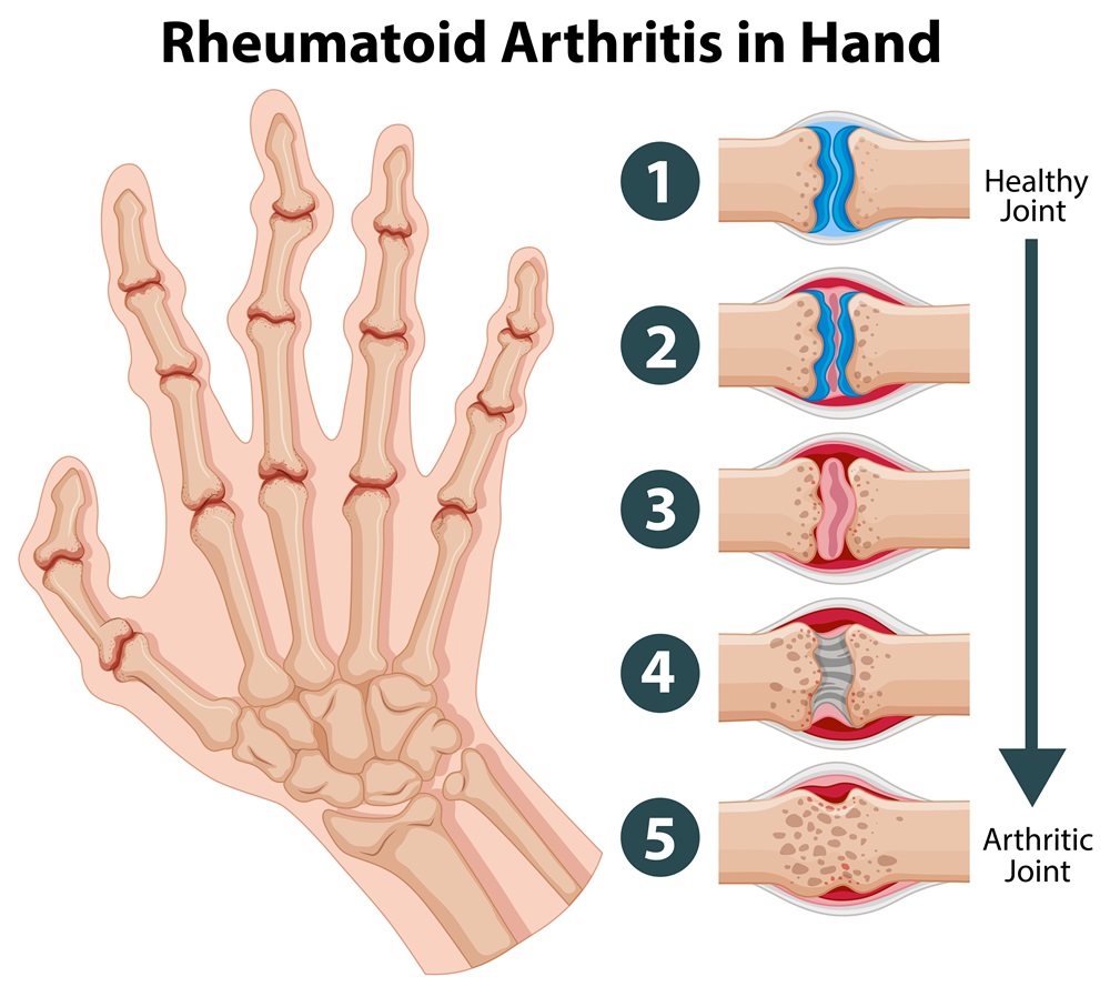
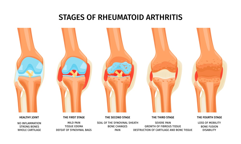
  • What are the symptoms of JIA in children?

Symptoms can vary, but common ones include swelling, persistent joint pain, morning stiffness, muscle weakness, and difficulty performing certain movements. It is important to note any unusual changes in your child’s behavior or activities.

  • How is JIA diagnosed in children?

The diagnosis of JIA often involves close collaboration between medical specialists, including pediatric rheumatologists. Medical examination, physical examinations, and blood tests can be used to confirm the diagnosis and rule out other causes of the symptoms.

  • What are the treatment options for children with JIA?

Treatment may include anti-inflammatory drugs, physical and occupational therapy, and in some cases, immune-modulating drugs. It is important to discuss the benefits and risks of each treatment option with your medical team.

  • What is involved in the daily management of JIA in the family?

For parents, day-to-day management of JIA may include taking medications regularly, attending physical or occupational therapy sessions, and closely monitoring symptoms. Open and honest communication with the child is also essential.

  • How does AIJ affect children’s school and social life?

JIA can bring additional challenges to children’s school and social life. Parents should work together with teachers and other professionals to ensure an appropriate and supportive learning and socializing environment.

  • What is the long-term outlook for children with JIA?

The long-term outlook varies depending on the severity of the disease. With appropriate treatment and effective management, many children with JIA can lead active and healthy lives. Regular medical monitoring is essential to detect and manage potential complications.

Frequently asked questions for parents

How can parents help their children?

For parents who have children diagnosed with Juvenile Idiopathic Arthritis (JIA), appropriate support and care play a critical role in managing the disease and ensuring the children’s quality of life. Here are some ways parents can help their children in this situation:

1. Education and Information

Understand as much as you can about JIA, treatment and symptom management. The more informed you are, the better informed decisions you will be able to make regarding your child’s health.

2. Open Communication

Establish open communication with your child. Encourage him to share his feelings and thoughts about the illness. An open relationship will make it easier to manage emotions and anxieties.

3. Involvement in Treatment

Actively participate in the treatment plan established by the medical team. Make sure medications are taken as prescribed and that recommended therapies are followed regularly.

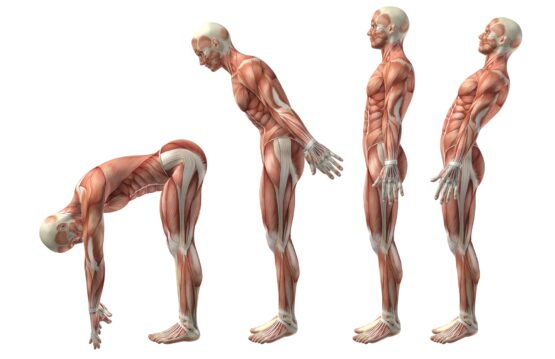
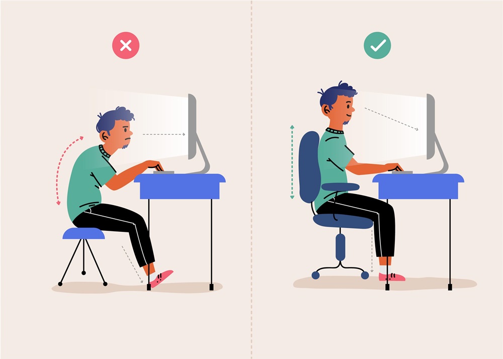
4. Promoting Healthy Lifestyle

Encourage a healthy lifestyle with an emphasis on a balanced diet and adequate physical activity. Ask the doctor about the right exercise for your child and create a program that fits his needs.

5. Monitoring Symptoms

Be aware of changes in your child’s symptoms and communicate this information to the doctor. Regular health monitoring will help to adjust the treatment plan according to the evolution of the disease.

6. Psychological Support

Provide emotional and psychological support to your child. It may also be helpful to involve counselors or therapists to provide additional assistance in managing emotions related to the illness.

7. Coordination with the School and Other Professionals

Communicate with teachers and school staff to ensure an appropriate learning environment for your child. Involve specialists, such as physical or occupational therapists, to support the child’s development in all aspects.

8. Encourage Autonomy

Encourage your child to be independent as much as possible. Give him opportunities to develop his skills and assume responsibilities within the limitations imposed by the illness.

9. Managing Family Stress

Every member of the family can be affected by JIA, so it is important to manage family stress. Find support in the community and share experiences with other parents who are going through similar situations.

Essentially, active involvement, emotional support and effective treatment management are the keys to helping children with Juvenile Idiopathic Arthritis lead as normal and healthy lives as possible.

How can parents help their children

Can juvenile arthritis be temporary?

Juvenile Idiopathic Arthritis (JIA) can have different evolutions depending on the specific form of the disease. In most cases, JIA is not a temporary condition and requires long-term management. However, there are also situations where the symptoms may disappear completely or be effectively controlled with treatment, allowing the child to lead an active and healthy life.

There are three main types of JIA, depending on the number of joints affected and other clinical characteristics:

  • Oligoarticular JIA (affects less than 5 joints): Some cases of oligoarticular JIA may go into remission, meaning complete disappearance of symptoms. However, it is important to note that in some cases the disease may recur or occur in other joints even after a period of remission.
  • Polyarticular JIA (affects more than 5 joints): This form can be persistent and requires long-term management. However, proper treatment can help control symptoms and prevent long-term complications.
  • Rheumatoid factor-negative and rheumatoid factor-positive JIA: These forms may have different courses and may require customized treatment strategies.

It is important to emphasize that the individual prognosis of a child with JIA can vary depending on several factors, including the specific form of the disease, the response to treatment, as well as the involvement of other organs in the inflammatory process. Early treatment and effective disease management can significantly improve the quality of life of children affected by Juvenile Idiopathic Arthritis. Always consult the doctor for evaluation and recommendations specific to your child.

Rheumatoid arthritis - 10 things you should know

Rheumatoid arthritis – 10 things you should know

Rheumatoid arthritis (RA) is a chronic, inflammatory condition that affects the joints, causing pain, swelling and stiffness. It is important to better understand this disease in order to be able to manage the symptoms and ensure a better quality of life for those affected. In this article, we will explore ten essential aspects related to rheumatoid arthritis.


CONTENT:

  1. RA causes
  2. Symptoms of RA
  3. Early diagnosis
  4. The Role of Genetic Factors
  5. Personalized Treatment
  6. Impact on Quality of Life
  7. Systemic complications
  8. The Role of Physical Exercises
  9. Alternative and Complementary Medicine
  10. Future Perspectives


1. RA causes

The causes of rheumatoid arthritis (RA) are complex and involve a combination of genetic and environmental factors. A central aspect of this condition is its autoimmune nature, where the immune system, responsible for defending the body against infection, misdirects its attention to its own tissues.

In rheumatoid arthritis, the immune system mistakenly attacks the synovial membranes, which are the tissues that cover the joints. These membranes are essential for the proper functioning of the joints, facilitating their movement and ensuring proper lubrication. When the immune system attacks these membranes, the result is inflammation, swelling and joint damage.

Although the exact mechanism that triggers this autoimmune reaction is not yet fully understood, there are several factors that may contribute to the onset of rheumatoid arthritis. An important factor is genetic predisposition. People with certain genes may have a higher risk of developing RA, but this is not the only cause.


2. Symptoms of RA

The symptoms of rheumatoid arthritis (RA) are diverse and can vary depending on the severity of the disease and how it progresses over time. It is important to recognize and understand these symptoms in order to initiate the earliest and most effective treatment. Here is a more detailed development of the characteristic symptoms of rheumatoid arthritis:

  • Joint Pain

Joint pain is a central symptom of rheumatoid arthritis and can affect several joints simultaneously or at different times during the course of the disease. This pain can be felt as a tingling, throbbing or burning sensation, and its intensity can range from mild to severe. The pain may be worse in the morning or after periods of inactivity.

  • Joint Swelling

Joint swelling is the result of inflammation produced by the immune system in the synovial membranes. Affected joints become swollen, tender to the touch and can sometimes have a higher temperature than the rest of the body. Persistent swelling can lead to joint damage and deformities.

  • Joint Stiffness

Joint stiffness is another hallmark of rheumatoid arthritis. Patients may experience joint stiffness, especially in the morning or after periods of inactivity. This symptom may affect the ability to move and may last more than an hour.

  • Muscle Weakness

Rheumatoid arthritis can cause muscle weakness, and patients may experience a general feeling of fatigue and exhaustion. This weakness can occur both in the muscles near the affected joints and throughout the body.

  • Symmetric Affection

A distinctive feature of rheumatoid arthritis is symmetrical joint damage. This means that usually the same joints on both sides of the body are involved at the same time. For example, if a finger joint on the left hand is affected, the same joint on the right hand is likely to show similar symptoms.

  • Variability of Symptoms

Rheumatoid arthritis symptoms can vary in intensity and may come and go during periods called “flare-ups” and “remissions”. Flare-ups are periods when symptoms worsen, while remissions are periods when symptoms are milder or even absent.

Symptoms of RA

3. Early diagnosis

Early diagnosis is an essential element in the effective treatment of rheumatoid arthritis (RA), having a significant impact on the prognosis and quality of life of patients. Here are some relevant points that emphasize the importance of early diagnosis in the management of rheumatoid arthritis:

  • Preventing Joint Damage

One of the main reasons early diagnosis is crucial in rheumatoid arthritis is the ability to prevent joint damage. In the early stages of the disease, prompt treatment can help control inflammation and slow down the process of joint tissue destruction. If rheumatoid arthritis is left untreated or is diagnosed at an advanced stage, irreversible damage to the joints can occur, which significantly affects the functionality and mobility of the patient.

  • Symptom Management and Quality of Life Improvement

Early diagnosis allows rapid initiation of an appropriate treatment plan, which can help control symptoms and improve the patient’s quality of life. Joint pain, swelling and stiffness can be managed more effectively with early therapeutic interventions, allowing the patient to maintain optimal joint function and lead as normal a life as possible.

  • Prevention of Systemic Complications

Rheumatoid arthritis doesn’t just affect the joints; it can also have systemic complications involving internal organs such as the heart, lungs, and blood vessels. Early diagnosis and appropriate management can help prevent these complications and maintain overall health.

  • Optimizing Treatment Response

Treatment of rheumatoid arthritis can vary depending on the stage of the disease and individual response to therapy. An early diagnosis gives the doctor the opportunity to more effectively adapt the treatment plan according to the specific needs of the patient and to optimize the management of the disease.

  • Patient Involvement in Therapeutic Decisions

Early diagnosis allows the patient to be more active in the decision-making process regarding treatment. Early understanding of the disease and treatment options gives the patient the opportunity to collaborate with the medical team in developing a personalized plan that aligns with their individual needs.

Early diagnosis

4. The Role of Genetic Factors

The role of genetic factors in the occurrence of rheumatoid arthritis (RA) is well established, and genetic predisposition is one of the main pillars of this autoimmune disease. However, it is important to emphasize that genetics is not the only determining factor in the development of AR, but interacts in a complex manner with environmental factors.

  • Genetic Predisposition

Studies have shown that there are certain genes associated with an increased risk of developing rheumatoid arthritis. HLA-DRB1 is an example of a gene with a strong link to AR. People who inherit certain variants of this gene have an increased risk of developing the disease. However, although genetic predisposition is important, not everyone who inherits these genes actually develops RA. Thus, environmental and other factors may intervene crucially.

  • Interaction with Environmental Factors

Even if a person has a genetic predisposition to RA, it is not an inevitable fate. Environmental factors, such as exposure to certain infections, smoking, stress and other lifestyle factors, can play an important role in triggering the disease in those with a genetic susceptibility. Infections, in particular, have been associated with the onset of rheumatoid arthritis in some cases, suggesting that they may activate the immune system and help initiate the autoimmune response.

  • The Complexity of Genetic and Environmental Interaction

The interplay between genetic and environmental factors in the occurrence of AR is complex and still not fully understood. It is believed that certain events or exposures in a person’s life can trigger abnormal immune reactions in those with a genetic predisposition. These incorrect immune reactions can cause the chronic inflammation characteristic of rheumatoid arthritis.

  • Importance of Risk Factor Assessment

Identifying the genetic and environmental factors associated with rheumatoid arthritis is essential to develop personalized prevention and treatment strategies. People with a family history of RA may benefit from closer monitoring and preventive interventions to minimize the risk of developing the disease.

The Role of Genetic Factors

5. Personalized Treatment

Management of rheumatoid arthritis usually involves a personalized approach. Anti-inflammatory medications, lifestyle changes, and physical therapy are just a few treatment options that can be customized to the individual patient’s needs.

6. Impact on Quality of Life

Rheumatoid arthritis can significantly affect the patient’s quality of life. Chronic pain and discomfort can lead to emotional disorders such as anxiety and depression. Emotional support and counseling can be essential in managing these issues.

7. Systemic complications

Rheumatoid arthritis (RA), in addition to its impact on the joints, can cause systemic complications, affecting internal organs and contributing to various conditions. Understanding these complications is crucial to ensure a complete and appropriate management of the disease.

  • Affecting the Cardiovascular System

Rheumatoid arthritis can increase the risk of cardiovascular disease. The chronic inflammation associated with RA can damage the artery wall, increasing the risk of atherosclerosis and blood clots. RA patients may also have a higher risk of heart disease and stroke. Blood pressure monitoring, cholesterol levels and prevention of cardiovascular risk factors thus become a priority.

  • Affecting the Respiratory System

The lungs can be affected in rheumatoid arthritis, causing complications such as pulmonary fibrosis, pleurisy or pneumonitis. These conditions can cause breathing difficulties and compromise the normal functioning of the lungs. Monitoring of respiratory function and periodic assessment of lung status are essential for early detection of these complications.

  • Affecting Blood Vessels

RA can affect blood vessels by causing vasculitis, an inflammation of the vessel wall. Vasculitis can affect various organs, including the skin, nerves, kidneys, and others. This complication can have serious consequences, and careful monitoring of the condition of the blood vessels is essential to prevent vascular complications.

  • Bone and Marrow Complications

Rheumatoid arthritis can contribute to bone loss and eventually osteoporosis. Weakened bones can increase the risk of fractures. Chronic inflammation can also affect the bone marrow, contributing to anemia or other hematological disorders. Bone density assessment and blood monitoring for abnormalities are necessary to prevent and manage these complications.

  • Eye Diseases

Rheumatoid arthritis can affect the eyes, leading to conditions such as Sjögren’s syndrome (an autoimmune disorder that affects the glands that produce tears and saliva) or scleritis (inflammation of the outer layer of the eye). Regular eye exams are essential for early detection and treatment of these problems.

Systemic complications

8. The Role of Physical Exercises

Regular exercise can play a crucial role in managing the symptoms of rheumatoid arthritis. They can improve patients’ mobility, muscle strength and mood.

9.Alternative and Complementary Medicine

Certain complementary therapies, such as acupuncture, massage, and yoga, can provide relief for rheumatoid arthritis. However, it is important to always talk to your doctor before trying such treatments.

10. Future Perspectives

Rheumatoid arthritis research is ongoing, and new therapies and approaches may bring hope for patients in the future. Involvement in clinical trials and maintaining open communication with the medical team can contribute to more effective disease management.

Surgical Treatment of Spinal Tumors

Surgical Treatment of Spinal Tumors: Exploring Options

Surgical treatment for spinal tumors navigates a complex, evolving frontier, uniting modern technology and medical expertise for innovative therapies. Spinal tumors, whether benign or malignant, pose significant challenges in their diagnosis and management.

In this context, surgical interventions often become the key to providing patients with not only symptomatic relief, but also improved quality of life prospects. In this regard, the detailed exploration of surgical methods, advanced technologies and advances in the treatment of spinal tumors brings to the fore a vital insight into this branch of neurological surgery.


CONTENT:

  1. Types of Spinal Tumors
  2. Indications for Surgical Treatment of Spinal Tumors
  3. Common Surgical Procedures
  4. Advanced Technologies and Minimally Invasive Approaches
  5. Potential Complications and Postoperative Management


Types of Spinal Tumors

In the complex world of spinal tumors, the diversity of these conditions requires a thorough understanding before considering surgical options. These tumors can be classified into various categories, each with distinct characteristics and implications for their management.

  • Meningiomas

Meningiomas are a category of tumors that develop from the membranes that cover the brain and spinal cord. They can vary in size and degree of malignancy, and the surgical approach depends on their location and extent.

  • Neuronomas

Neurinomas, also known as schwannomas, are tumors that develop around nerves and can affect the spinal cord. The surgical approach focuses on preserving the nerves and minimizing the risks to their functionality.

  • Cancer Metastases

Metastatic spinal tumors come from cancer that has spread from other areas of the body. They can affect the vertebrae and spinal cord, often requiring an integrated approach, including surgery to relieve pressure and adjuvant therapies to manage the primary cancer.

  • Glioblastomas

Glioblastomas are aggressive brain tumors that, in some cases, can spread to the spinal cord. Surgery may be needed to remove or shrink the tumor mass, although treatment often also includes radiation therapy and chemotherapy.

Each type of spinal tumor presents unique challenges, and the treatment plan must be tailored to the specifics of each case. Before approaching surgical treatment, a detailed assessment of the type of tumor, its stage and the impact on the patient is essential to ensure an effective and personalized approach.

Types of Spinal Tumors

Indications for Surgical Treatment of Spinal Tumors

The decision to resort to surgical treatment for a spinal tumor is a complex process, influenced by a number of factors that must be carefully evaluated. These essential factors determine the nature and extent of surgery, adapting to the specific needs of each patient.

1. Tumor size

The size of the tumor is an important criterion in determining the need for surgery. Large tumors can exert significant pressure on adjacent structures, causing symptoms and affecting quality of life. In these cases, the surgical approach may be essential to reduce the tumor mass and restore the normal function of the spine.

2. Tumor location

The location of the tumor is another determining factor in the decision to opt for surgical treatment. Tumors located near the spinal cord or nerves can create severe complications, which is why surgery may be considered to minimize risks and preserve nerve functionality.

3. Degree of Malignancy

The degree of malignancy of the tumor is crucial in defining the treatment strategy. Malignant tumors, which have an increased potential for spread and recurrence, may require a more aggressive surgical approach, often associated with complementary therapies such as radiotherapy or chemotherapy.

4. Symptoms Presented by the Patient

The symptoms experienced by the patient play a central role in making the decision for surgical treatment. Persistent pain, weakness, difficulty moving, or changes in pelvic organ function may indicate the need for surgery to relieve or prevent progression of symptoms and improve the patient’s quality of life.

By carefully evaluating these factors, the medical team can create a personalized treatment plan, adapted to the specifics of each case. The decision to choose surgical treatment for a spinal tumor reflects a balance between effectively removing the tumor and minimizing the impact on spinal and nervous system functionality.


Common Surgical Procedures

1. Tumor Resection:

Tumor resection is one of the fundamental surgical interventions in the treatment of benign spinal tumors. This procedure aims at the complete excision of the tumor mass, thus eliminating any risk of recurrence and restoring the integrity of the affected tissues. Usually performed for tumors with benign features, tumor resection aims to preserve the health of the spine and the functionality of the spinal cord.

2. Spinal Cord Decompression:

Tumors that grow near the spinal cord can put pressure on it, causing symptoms such as pain, weakness, or difficulty moving. The spinal cord decompression procedure involves removing or reducing the pressure exerted by the tumor on the nerve structures. This intervention not only relieves symptoms, but also prevents further damage to the spinal cord, thus contributing to improving the patient’s quality of life.

3. Stabilization and Reconstruction:

In situations where the tumors are malignant or affect the bony structures of the spine, surgical procedures for stabilization and reconstruction become the imperative of treatment. These interventions have the role of strengthening the spine, preventing instability and subsequent deformations. Reconstruction also focuses on restoring the integrity of the affected bones and neighboring structures, thus maintaining the stability of the spine and its functionality.

Common Surgical Procedures

Advanced Technologies and Minimally Invasive Approaches

A significant evolution in the field of spinal tumor surgery has been represented by advanced technologies and minimally invasive approaches. Advanced imaging such as magnetic resonance imaging (MRI) and computed tomography (CT) provides surgeons with detailed information for precise intervention planning.

Minimally invasive approaches such as laser surgery and endoscopic techniques have the advantage of reducing tissue trauma and postoperative recovery time. These methods allow, in many cases, to perform the surgical intervention with smaller incisions, without compromising the quality of the treatment.


Potential Complications and Postoperative Management

Regardless of the nature of the surgery, the treatment of spinal tumors comes with risks and potential complications that require a careful and anticipatory approach. These complications include:

  • Bleeding

Excessive bleeding during or after surgery can be a serious complication, requiring immediate management to prevent further complications.

  • Infections

Postoperative infections are a risk given the opening of tissues and exposure to potentially contaminating environments. Correct administration of antibiotics and close monitoring are essential in preventing and treating infections.

  • Nerve Damage

Surgery near nerves may cause damage. They can cause temporary or permanent loss of nerve functionality, affecting the patient’s quality of life.

  • Affecting the Functionality of the Spinal Cord

Especially with spinal cord-adjacent tumors, there’s a risk of post-surgery functional impact. Moreover, careful control of this aspect is crucial to avoid severe complications.

  • Postoperative Management

After surgery, postoperative management plays a vital role in ensuring an optimal recovery for the patient. It involves:

1. Physiotherapy

The personalized physiotherapy program helps to restore muscle strength, mobility and functionality. Exercises are tailored to minimize the risk of complications and support spinal recovery.

2. Occupational Therapy

Occupational therapy focuses on restoring independence and the ability to perform daily activities. This improves the patient’s quality of life and contributes to reintegration into society.

3. Adjuvant Therapy

In some cases, adjuvant therapies such as radiotherapy or chemotherapy may be necessary to complete the treatment and prevent relapses.

However, medical teams aim to optimize patient recovery and support a return to an active, healthy life through meticulous complication management and thorough postoperative care.

Kyphosis treatment News and innovations

Kyphosis treatment: News and innovations

Kyphosis treatment is an ever-evolving field, where scientific and technological advances are opening new horizons in addressing this spinal condition. With constant research and discoveries, medical specialists have introduced innovative methods into the therapeutic arsenal of kyphosis aimed at optimizing treatment results.

In this context, we explore in detail the novelties and advances in the field of treatment for kyphosis, highlighting the modalities that are redefining the perspective on the management of this medical condition.


CONTENT:

  1. History of Kyphosis Treatment
  2. New methods of treatment
  3. Exaggerated kyphosis treatment


History of Kyphosis Treatment

The history of kyphosis treatment reflects the continuous evolution of the methods and technologies used to address this complex spinal condition. In the past, kyphosis therapy focused on traditional methods, such as physical therapy and specific exercises, mainly aimed at strengthening the back and abdominal muscles. These approaches were designed to improve posture and prevent excessive curvature of the spine from worsening.

Posture-correcting devices such as corsets or braces have also been used to provide additional support and encourage proper spinal alignment. In more severe cases, where the spinal deformity was pronounced and caused significant complications, surgery was a viable option. These procedures focused on physically correcting the spine to restore proper alignment and reduce pressure on the intervertebral discs and nerves.

However, technological advances and scientific research in recent decades have opened new perspectives on the treatment of kyphosis. New discoveries in science and technology have enabled the development of more advanced and personalized approaches. Exoskeletons, for example, have become a promising innovation, providing personalized biomechanical support and helping to maintain correct posture during daily activities.

In addition, technologies such as virtual reality and artificial intelligence have been integrated into the treatment process, providing interactive and personalized methods for addressing kyphosis. These tools allow therapists to create exercise programs tailored to individual patient needs and monitor progress in more detail.

History of Kyphosis Treatment


New methods of treatment

In the medical field, rapid advances and innovative discoveries have opened up new horizons for addressing ailments and improving patients’ quality of life. A significant aspect of this revolution is the development and implementation of new treatment methods, which bring significant benefits in the management of various health conditions. These methods represent not only an evolution of traditional medical practices, but also an adaptation to the complex challenges of contemporary society.

New therapeutic approaches meet the individual needs of patients, taking into account the variability of genetic factors, lifestyles and social contexts. Recent breakthroughs in biotechnology, artificial intelligence and personalized medicine have contributed to the development of a dynamic medical landscape, where traditional methods are complemented or even replaced by more precise and effective interventions.

1. Exoskeleton therapy

One of the most recent advances in the treatment of kyphosis is the use of exoskeletons. These portable devices, which attach directly to the body, are designed to support and correct the patient’s posture. People can customize them to suit their individual needs and use them during daily activities to maintain correct posture.

2. Virtual Reality (VR) Therapy

The use of virtual reality in the treatment of kyphosis has become increasingly popular. Patients can perform specific exercises in a virtual environment under the close supervision of the therapist. This type of therapy not only provides an interactive and fun aspect, but also helps improve the patient’s motivation to follow a regular treatment schedule.

3. Gene therapy

In medical research, gene therapy has become a growing area of interest. In the case of kyphosis, researchers are exploring the possibility of using gene therapy to correct genetic abnormalities associated with the development of this condition. Although this field is in the early stages of research, initial results show promise in the direction of developing more specific treatments.

4. Artificial Intelligence (AI) Therapy

The use of artificial intelligence in the diagnosis and treatment of kyphosis is another significant innovation. Machine learning algorithms can analyze data from medical images and provide personalized recommendations for treatment. This type of approach can improve diagnostic accuracy and adapt therapy to individual patient changes over time.

New methods of treatment

Exaggerated kyphosis treatment

Treatment of exaggerated kyphosis depends on the severity of the condition, the patient’s age, general health and possible associated complications. The treatment includes physical therapy, exercises, posture correction, and, in some cases, medical or surgical interventions. It is crucial to see a medical specialist, such as an orthopedist or physical therapist, for an evaluation and personalized treatment plan. Here are some common treatment methods for exaggerated kyphosis:

1. Physical Therapy and Exercise:

  • Physiotherapy can help strengthen your back and abdominal muscles, thereby improving your posture.
  • Specialized exercises focused on strengthening the muscles around the spine can help reduce excessive curvature.

2. Posture Correction:

  • Education about proper posture during daily activities and at the office can help prevent kyphosis from worsening.
  • Medical professionals may recommend postural supports, such as corsets or braces, to help maintain proper spinal alignment.

3. Drug Therapy:

  • Doctors may prescribe nonsteroidal anti-inflammatory drugs (NSAIDs) to control pain associated with kyphosis.
  • In some cases, doctors may use muscle relaxant medications to reduce muscle spasms.

4. Medical Interventions:

  • Physicians may administer corticosteroid injections to reduce inflammation around the spine and relieve pain.
  • Therapy with the help of a pain specialist may also involve techniques such as nerve ablation to reduce pain sensations.

5. Surgery:

  • In severe cases or when other treatment methods prove unsuccessful, physicians may consider surgery to correct the spinal deformity.

It is essential to address exaggerated kyphosis under the guidance of a medical professional and follow a personalized treatment plan. Self-treatment or ignoring symptoms can lead to complications and progression of the condition.

Chiropractic Treatment for Spondylolisthesis An Effective Approach

Chiropractic Treatment for Spondylolisthesis: An Effective Approach

Chiropractic treatment has proven to be an effective and holistic option for managing spondylolisthesis, a spinal condition that can cause pain and discomfort.

Spondylolisthesis is characterized by the abnormal sliding of one vertebra over another, and chiropractic therapy offers a non-invasive alternative to conventional treatments. In this article, we will explore how chiropractic can help relieve symptoms and improve the quality of life for those affected by spondylolisthesis.


CONTENT:

  1. How does chiropractic treatment work?
  2. Initial assessment and treatment plan
  3. Chiropractic adjustments and symptom reduction
  4. Exercises and recommendations for self-care


How does chiropractic treatment work?

Chiropractic treatment is based on the principle that a correctly aligned spine contributes to the optimal functioning of the entire nervous system, and implicitly, to the overall health of the body. The chiropractor’s primary concern is to identify and correct structural imbalances in the spine that may affect the normal flow of information between the brain and the rest of the body.

  • Correct alignment of the vertebrae

Essentially, the spine serves as the central axis of the human body, and therefore any displacement or misalignment of the vertebrae can affect the entire nervous system. Chiropractors use manual manipulation techniques to return the vertebrae to their correct position. This process, called a chiropractic adjustment, involves applying controlled and directed pressure to specific parts of the spine or joints to restore proper alignment.

  • Reducing nerve pressure

Spondylolisthesis can lead to increased pressure on nerves, causing pain and discomfort. Chiropractic adjustments aim to reduce this pressure. This is done by correcting the position of the vertebrae, thus removing any constraint or compression on the nerves. By reducing nerve pressure, chiropractors help improve the transmission of nerve signals and restore normal body functions.

  • Stimulation of natural healing processes

Another component of chiropractic treatment is the stimulation of the body’s natural healing processes. Chiropractic adjustments can improve blood circulation and promote the release of endorphins, the body’s natural chemicals that reduce pain and improve well-being. This holistic approach helps speed up the recovery process and strengthen the body’s ability to heal itself.

  • Treatment customization

Each patient has individual needs and conditions, and the chiropractic approach adapts according to these particularities. Chiropractors consider the patient’s medical history, specific symptoms, and physical assessment to develop a personalized treatment plan. This plan may include various techniques and exercises to optimize the recovery process.

How does chiropractic treatment work

Initial assessment and treatment plan

In chiropractic treatment, the initial assessment is an essential pillar for understanding the specific needs of the patient and developing an effective treatment plan. The chiropractor begins with a detailed discussion of the patient’s medical history, investigating any previous conditions, treatments followed and relevant health details. This conversation provides a comprehensive picture of the patient’s medical background and provides crucial information for developing a tailored treatment plan.

Development of the treatment plan

Based on the information obtained from the initial assessment, the chiropractor develops a personalized treatment plan. This plan is tailored to the patient’s individual needs and aims to correct the problems identified during the assessment. The main elements of the treatment plan include:

  • Regular manual adjustments: The chiropractor uses manual manipulation techniques to adjust the spine and joints, helping to restore proper alignment and reduce nerve pressure.
  • Strengthening exercises: The patient can be recommended specific exercises to strengthen the muscles around the spine, thus supporting the healing process and preventing recurrence.
  • Pain Management Techniques: The chiropractor may introduce pain management techniques, including ice/heat therapy or relaxation techniques, to relieve discomfort and improve the patient’s quality of life.
  • Follow-up schedule: The treatment plan may include regular scheduling for adjustments and periodic assessments to monitor progress and adapt interventions as the patient’s condition evolves.

Initial assessment and treatment plan

Chiropractic adjustments and symptom reduction

1. Chiropractic Adjustments – Precise Alignment Technique

Chiropractic adjustments are the core of chiropractic therapeutic intervention and are essential in treating spondylolisthesis. Furthermore, these adjustments involve the use of precise and controlled manual movements applied to the vertebrae and joints. In the case of spondylolisthesis, where one vertebra slips in front of the one below, these adjustments are meant to correct the incorrect position and restore normal spinal alignment.

2. Reduction of vertebral slippage

Spondylolisthesis causes the vertebrae to slip abnormally, which can lead to nerve compression and pain. Moreover, chiropractic adjustments target exactly this problem, providing a non-invasive solution to reduce vertebral slippage. The chiropractor applies specific pressures to guide the vertebra back into the correct position, thereby eliminating tension and excessive pressure on the nerves.

3. Relief of nerve pressure

Excessive pressure on the nerves due to spondylolisthesis can cause pain and discomfort. Chiropractic adjustments not only correct spinal alignments, but also reduce associated nerve pressure. By removing nerve compression, the chiropractor facilitates the transmission of nerve signals, thereby helping to reduce symptoms such as pain, tingling or weakness in the affected areas.

4. Patient experience

Many spondylolisthesis patients report significant improvement after regular chiropractic treatment sessions. They notice a reduction in pain and an increase in mobility in daily activities. Chiropractic adjustments can help restore a normal state of the spine, allowing patients to once again enjoy an increased quality of life and regain control of their physical functions.

5. Continuation of treatment and prevention of relapses

Chiropractic adjustments can have an immediate impact on symptoms, but it is important to recognize that treating spondylolisthesis requires ongoing commitment. The chiropractor will develop a long-term treatment plan that may include regular adjustments, strengthening exercises, and education to properly manage your posture and daily activities. This holistic approach not only relieves existing symptoms, but also helps prevent relapses and maintain a healthy spine in the long term.

Exercises and recommendations for self-care

Exercises and recommendations for self-care

In the treatment of spondylolisthesis, muscle strengthening exercises are an essential element of the treatment plan. Chiropractors can develop a personalized set of exercises tailored to the patient’s specific needs and fitness level. These exercises aim to strengthen the muscles around the spine. Thus, the affected area is stabilized and unwanted movements of the vertebrae are prevented.

  • Spinal Stabilization Exercises

To strengthen support for the spine, patients may be instructed to perform exercises that focus on stabilizing the spine. Stabilization exercises improve muscle strength and control around the vertebrae, helping to maintain proper alignment and prevent relapses. Additionally, these may include exercises with fitness balls, planks, or other movements that emphasize the involvement of core muscles.

  • Self-care and prevention techniques

In addition to exercise, chiropractors offer advice to patients on self-care and prevention techniques. These may include:

1. Posture correction: Educating patients about correct posture is crucial. Chiropractors offer practical advice for maintaining balanced posture during daily activities and at work.

2. Muscle relaxation techniques: Chiropractors can train patients in muscle relaxation techniques, which can reduce tension and discomfort in the spinal area.

3. Managing stress: Stress can contribute to muscle tension and deterioration of spinal health. Recommendations for managing stress may include deep breathing techniques, meditation, or other relaxation methods.

4. Monitoring physical activities: Chiropractors can advise patients on appropriate physical activities and how to avoid movements that may aggravate spondylolisthesis. Special attention is paid to how patients lift objects or carry out their daily activities.

  • The Importance of Continuity in Self-Care

Self-care exercises and techniques are often an essential part of a long-term maintenance regimen. Continuing exercises and self-care post-symptom improvement reinforces results and prevents relapses in spondylolisthesis. Patients should incorporate these practices for a healthy spine and to sustain the long-term benefits of chiropractic treatment.

Laminectomy for lumbar stenosis

Lumbar laminectomy for spinal stenosis – Effective Surgical Option

Lumbar laminectomy is revealed as an essential answer for those experiencing spinal stenosis, a painful condition that affects the spinal canal in the lower region of the spine.

Spinal stenosis, characterized by the narrowing of this vital canal, can cause a variety of symptoms, including persistent lower back pain and discomfort in the lower limbs. In this context, lumbar laminectomy is an advanced and effective surgical approach designed to provide patients with a path to relief and restoration of quality of life.


CONTENT:

  1. What is Lumbar Laminectomy?
  2. Performing the Procedure: Step by Step
  3. Recovery and Benefits of Lumbar Laminectomy
  4. Conclusion: A Solution with Bright Prospects


What is Lumbar Laminectomy?

Lumbar laminectomy is a surgical procedure that involves removing a portion of the bones called the laminae from the back of the lumbar vertebrae. This strategic intervention is primarily aimed at creating additional space in the spinal canal, removing pressure on the nerves and spinal cord. In essence, laminectomy paves the way for these delicate structures to regain their freedom and function within normal parameters.

Lumbar laminectomy for spinal stenosis - Effective Surgical Option

Performing the Procedure: Step by Step

1. Preoperative Assessment: Confirming and Elaborating the Diagnosis

Before starting the intervention, the patient undergoes a thorough preoperative assessment. This includes laboratory tests, advanced medical imaging such as computed tomography or magnetic resonance imaging, and a detailed discussion with the specialist. The purpose of this stage is to confirm the diagnosis of spinal stenosis, to identify the anatomical peculiarities of the affected region and to establish a personalized surgical plan.

2. Anesthesia: A State of Comfort for the Patient

After the preoperative assessment is complete, the patient enters the operating room, where general anesthesia is administered. This procedure ensures that the patient is completely asleep and does not feel pain during the operation. Maintaining a comfortable position throughout the procedure is essential for as smooth a recovery as possible.

Anesthesia

3. Access to the Spine: Surgical Incision

With the patient under general anesthesia, the surgeon begins the procedure by making an incision in the lumbar region. Surgeons make this incision precisely to allow access to the affected vertebrae and ensure adequate visualization of the intervention area.

4. Removing the Slats: Creating the Necessary Space

With access secured, the surgeon uses specialized instruments to remove small segments of bone called lamellae. This step is crucial for creating additional space in the spinal canal, thereby eliminating excessive pressure on the nerves and spinal cord.

5. Nerve Release: Reducing Pressure and Correcting Abnormality

Removing the lamellae allows the surgeon to identify and correct any pathological tissue changes that may be contributing to the spinal stenosis. The pressure on the nerves and spinal cord is significantly reduced, facilitating the recovery of these sensitive structures.

6. Closing the Incision: Toward Completion of the Procedure

At the end of the lumbar laminectomy, the surgeon carefully closes the incision with surgical sutures. Furthermore, this step is essential to ensure proper wound healing and to minimize the risk of infection.

Through this meticulous, step-by-step approach, lumbar laminectomy becomes an effective surgical option for patients seeking relief from spinal stenosis. Carefully and precisely carrying out each step of the procedure is important to ensure optimal results and a recovery as fast and comfortable as possible.

Recovery and Benefits of Lumbar Laminectomy

After the lumbar laminectomy is completed, a crucial stage of the medical process begins: the patient’s recovery. This period can vary depending on the complexity of the surgery and the health of each individual, but the tangible benefits of laminectomy often become apparent during and after this recovery phase.

Recovery from a lumbar laminectomy can take an average of 4 to 6 weeks, but the exact time can vary depending on the complexity of the procedure and the individual patient’s progress.

After such surgery, patients can start walking quite quickly, often even in the first days after the operation. However, returning to normal activities and walking without support may take several weeks.

  • Improvement of Symptoms

Many patients experience significant improvement in symptoms immediately after surgery. Lumbar pains that were initially present may diminish or even disappear, and sensations of tingling or weakness in the lower limbs may experience a considerable improvement. This development brings a sense of relief and comfort, giving the patient a much improved quality of life.

  • The role of Physiotherapy

Physiotherapy often becomes an essential component of the recovery process. Specialized exercises and techniques guide patients to strengthen the muscles around the spine, improve posture, and regain flexibility. These physiotherapy sessions aim to reinforce the results of the surgical procedure, ensuring a full recovery and preventing the recurrence of symptoms in the future.

  • Returning to Moderate Activities

As the patient progresses in recovery, it is recommended to gradually resume moderate activities. Even though rest is essential for the first few weeks, once the doctor deems it safe, the patient can gradually return to normal daily activities. This can include walking, swimming, or other light physical activity that supports spinal strength and flexibility.

  • Postoperative monitoring

After the back operation, it’s crucial to keep checking the patient’s health regularly. Additionally, seeing the doctor regularly helps check how well you’re recovering and adjust any advice about exercise or treatment.

  • Improving the Quality of Life

Over time, lumbar laminectomy helps improve the quality of life. It allows patients to move better and do daily tasks without ongoing pain and discomfort, regaining their independence.

Recovery

Conclusion: A Solution with Bright Prospects

Lumbar laminectomy is emerging as an effective surgical option, offering relief and hope to patients affected by spinal stenosis. However, decide after discussing with your specialist, considering benefits, and respecting risks. In light of medical advances, lumbar laminectomy is thus becoming a gateway to an improved quality of life for those dealing with this complex condition.


Vertebral Fractures: Types and a detailed look

Vertebral Fractures: Types and a detailed look

Vertebral fractures represent significant medical events, characterized by damage to the bone structures that make up the spine. This region of the body has an essential role in maintaining stability and mobility, and fractures in this area can have serious consequences for the patient’s health. There are various causes that can contribute to these fractures, including trauma, conditions such as osteoporosis, or other pre-existing medical conditions.

Trauma and Causes of Vertebral Fractures

A common factor that can lead to vertebral fractures is trauma, resulting from events such as car accidents, severe falls, or sports accidents. Sudden force applied to the spine can cause fractures of varying degrees of severity.

Osteoporosis and Bone Fragility

Another important cause is osteoporosis, a medical condition characterized by loss of bone density. In osteoporosis, the bones become more fragile and susceptible to fractures. Vertebral fractures in this category can occur from even minor trauma or ordinary pressure.

Other Contributing Medical Conditions

There are other medical conditions, such as bone tumors or spinal infections, that can contribute to fractures. These conditions can weaken the bone structure or exert excessive pressure on them, favoring fractures.

In this context, the article aims to provide a detailed insight into the different types of vertebral fractures, understanding the specific symptoms associated with each type and the available treatment modalities. In addition to this aspect, it is crucial to also examine the potential complications that can occur following vertebral fractures, in order to fully understand the impact on the patient’s health and to guide the recovery process towards an appropriate and effective approach.


CONTENT:

  1. Compression Vertebral Fractures
  2. Breakage Fractures
  3. Flexion fractures
  4. Settlement Vertebral Fractures
  5. Fractures in Bulk


Compression Vertebral Fractures

Compression fractures are a specific type of spinal injury that occurs when a vertebra is subjected to excessive pressure, usually from a fall or direct impact. This form of fracture is distinguished by the way it affects the morphology of the respective vertebra.

  • Causes and Triggers

Compression fractures are often the result of a vertical force applied rapidly to the spine. Falls from heights or direct impact, such as in an accident, can create concentrated pressure on one or more vertebrae.

  • Fracture Mechanism

In the case of compression fractures, the vertebra undergoes significant pressure, causing excessive compression of the bone. This process leads to a change in the shape and size of the affected vertebra, usually resulting in a shorter and wider configuration than normal.

  • Characteristic symptoms

Symptoms associated with compression fractures include intense local pain in the affected area. Patients may experience discomfort or pain in the spine, especially around the affected vertebra. Another frequent manifestation is the adoption of a bent or bent posture, reflecting the structural changes of the vertebra.

  • Diagnosis and Treatment

Diagnosing compression fractures often involves medical imaging, such as X-rays or CT scans, to accurately assess the extent of the injury. Treatment may vary depending on the severity of the fracture and may include immobilization with braces, pain medication to manage pain, and, in some cases, physical therapy to maintain or restore mobility.

Compression Vertebral Fractures

Breakage Fractures

Burst fractures are a distinct type of spinal injury in which the bone is split into two separate parts. This type of fracture can involve significant damage to the bone structure and is often associated with severe trauma, such as car accidents or falls from a height. Exploring the characteristics of these fractures is essential to understanding the consequences and appropriate treatment modalities.

  • Causes and Traumatic Context

Breakage fractures are usually caused by traumatic events such as car accidents, severe falls, or other high-impact incidents. Sudden force applied to the spine can cause the bone to split into two distinct parts, placing considerable stress on the integrity of the bone structure.

  • Sharp Pain and Characteristic Symptoms

Patients with comminuted fractures often experience intense, sharp pain in the affected area. This pain may be localized around the affected vertebra and may radiate to other parts of the spine or body. In addition to pain, patients may experience significant difficulty moving the spine due to the severe injury.

  • Numbness and Weakness in the Lower Limbs

Another common symptom associated with comminuted fractures is numbness or weakness in the lower limbs. This is due to compression or damage to the nerves near the fracture, affecting the proper transmission of nerve signals and muscle strength in that area.

  • Diagnosis and Treatment

The diagnosis of comminuted fractures often involves the use of advanced imaging techniques such as computed tomography or magnetic resonance imaging. Treatment may involve immobilizing the spine with braces, pain medication to control pain, and, in some cases, surgery to restore bone structure.

Flexion fractures

Flexion fractures are injuries to the spine that occur when the spine is subjected to excessive bending or flexion. This type of fracture can have serious consequences on the bone structure and functionality of the spine, and is often associated with events such as a sudden impact or a fall.

  • Causes and Background of the Event

Flexion fractures are often caused by a quick impact or sudden force applied to the spine, causing it to bend excessively. Falls, sports accidents, or any other incident that generates considerable force on the vertical axis of the spine can contribute to this type of fracture.

  • Painful Symptoms and Difficulty in Movement

Localized pain in the affected area of the spine is a primary symptom of flexion fractures. The pain can be intense and radiate to other parts of the body. Patients may also experience significant difficulty moving the spine, which can affect the ability to perform normal daily activities.

  • Affecting Internal Organs

In severe cases, flexion fractures can affect the function of internal organs. The spine protects the spinal cord and plays an essential role in maintaining the integrity of the central nervous system. A severe flexion fracture can compress the spinal cord or damage the nerves that control organ functions, causing additional complications.

  • Diagnosis and Therapeutic Approach

The diagnosis of flexion fractures usually involves the use of medical imaging, such as X-rays or CT scans, to assess the extent and location of the injury. Treatment can vary depending on the severity of the fracture, including immobilizing the spine with braces or casts, pain medication to manage pain, and in some cases, surgery to restore proper alignment of the vertebrae.

  • Rehabilitation and Recovery Process

The recovery process often involves physical therapy aimed at restoring mobility, muscle strength, and normal spinal function. Careful monitoring of potential complications, such as nerve damage or internal organ dysfunction, is crucial for the long-term management of patients.

Flexion fractures


Settlement Vertebral Fractures

Injuries to the spine occur when an axial force is applied directly to it, causing compression fractures. This type of fracture characterizes the sudden compression or settling of the vertebrae, resulting in a significant impact on the structure of the spine. These fractures are common in high-impact accident situations, such as falls from heights or vehicle accidents.

  • Axial Forces and Fracture Mechanism

Axial force applied directly to the axis of the spine causes compression fractures. This type of force can occur at the time of an impact, such as a fall from a height or a vehicular collision. The applied force suddenly compresses and flattens the vertebrae.

  • Causes and Context of Traumatic Events

Traumatic events such as severe falls, car accidents, or falls from a height often associate with dislocation fractures. The violent impact of these events can put extreme pressure on the spine, causing compression fractures.

  • Variation of Symptoms

Symptoms of compression fractures can vary depending on the severity of the injury. Severe pain in the affected area is common, and patients may experience difficulty in moving the spine. In more severe cases, these fractures can affect surrounding structures, causing difficulty breathing or other respiratory problems.

  • Diagnosis and Medical Imaging

The diagnosis of subsidence fractures usually involves the use of advanced imaging techniques such as computed tomography or magnetic resonance imaging. These procedures allow doctors to assess the extent and severity of the injury, providing crucial information for treatment planning.

  • Management and Treatment

The treatment of compression fractures may involve immobilizing the spine with the help of corsets or specific devices. Proper pain management and management of complications, such as impaired breathing, are critical aspects of the recovery process.

  • Recovery and Long-Term Monitoring

The recovery process for compression fractures may require physical therapy to restore mobility and muscle strength. Closely monitor patients long-term to detect any potential complications and ensure as complete a recovery as possible.

Settlement Vertebral Fractures

Fractures in Bulk

Bulk fractures represent a category of spinal injuries characterized by simultaneous damage to several vertebrae. These fractures occur in extreme impact situations, such as plane crashes or falls from great heights, and can have severe consequences on the patient’s health and functionality. Detailed exploration of these injuries is crucial to understanding their complexity and implication in healthcare.

  • Extreme Impact and Bulk Fracture Mechanism

Bulk fractures occur in extreme impact circumstances characterized by strong forces applied to the spine. Airplane crashes, falls from great heights, or other high-energy incidents can cause simultaneous damage to multiple vertebrae.

  • Symptoms Severity of Bulk Fractures

Symptoms associated with bulk fractures are often severe and can vary depending on the severity of the injuries. Intense pain in the affected area is common and can be difficult to control with regular medications. Loss of motor function is a major concern, and patients may experience significant difficulty in performing daily movements.

  • Paralysis and Serious Complications

In extreme cases of bulk fractures, paralysis can occur as a result of damage to the spinal cord. This can affect the functionality of the lower or upper limbs, having a significant impact on the patient’s quality of life. Complications can also include bladder and bowel control problems, often requiring specialist medical attention and intensive rehabilitation.

  • Diagnosis and Prompt Intervention

The diagnosis of bulk fractures often requires the use of advanced imaging techniques, such as computed tomography or magnetic resonance imaging, to assess the extent and impact of the injuries. Prompt medical intervention is essential to stabilize the spine and minimize the risk of complications.

  • Therapeutic Approach and Rehabilitation

Treatment of bulk fractures can involve complex surgery to realign and fix the affected vertebrae. Immobilization of the spine with specific devices, physical therapy and rehabilitation are essential components of the recovery process, aiming to restore functionality and reduce the risk of long-term complications.

The Impact of Spinal Cord Lesions on the Urinary Bladder

Spinal Cord Injuries and the Impact on Bladder Function

Spinal cord injuries are a complex and often devastating medical problem, with the potential to radically change a person’s quality of life. The spinal cord is responsible for transmitting nerve impulses between the brain and the rest of the body, and any damage to this essential structure can have significant consequences.

A spinal cord injury can profoundly affect one of the fundamental aspects of daily life: bladder function. This vital function of the body, usually carried out involuntarily and under nervous control, often becomes a challenge for those with spinal cord injuries. Understanding how these lesions influence bladder health and identifying effective ways to manage them become essential in ensuring an optimal quality of life for these patients.

In this article, we’ll explore how spinal cord injuries can affect bladder health, as well as options for managing bladder problems.


CONTENT:

  1. Anatomy and Function of the Spinal Cord
  2. The Impact of Spinal Cord Lesions on the Urinary Bladder
  3. How can this problem be treated?


Anatomy and Function of the Spinal Cord

The spinal cord is a crucial structure that runs along the spine and is responsible for transmitting nerve impulses between the brain and the rest of the body. These impulses play a vital role in the control and coordination of movements and sensations. When an injury occurs in this area, it can significantly affect the body’s normal functions.

Anatomy and Function of the Spinal Cord

The Impact of Spinal Cord Lesions on the Urinary Bladder

The impact of spinal cord injuries on the urinary bladder is profound and can generate a number of complex problems related to the control and functioning of this vital organ. The spinal cord plays an essential role in transmitting the nerve signals that control bladder activity, and any injury to this area can disrupt the delicate coordination of the urination process.

1. Bladder Control

  • Bladder Muscle Contractions: The spinal cord is involved in the transmission of nerve signals that coordinate bladder muscle contractions. These contractions are essential to squeeze and eliminate urine effectively.
  • Function of the Sphincters: The sphincters, the muscles at the mouth of the bladder and urethra, are responsible for controlling the flow of urine. The spinal cord regulates the opening and closing of these sphincters in a coordinated manner to allow or stop the elimination of urine.

2. Fine Coordination Disorder

Spinal cord injuries can disrupt this delicate coordination between the spinal cord and the bladder, with the following consequences:

  • Urinary Retention: Lesions can lead to difficulty emptying the bladder completely. Lack of proper coordination between the spinal cord and the bladder can cause the bladder muscles to not contract properly, causing urinary retention.
  • Urinary Incontinence: On the other hand, injuries can affect the control of the sphincters, leading to urinary incontinence. Inability to keep the sphincters properly closed can lead to involuntary leakage of urine.
  • Recurrent Urinary Tract Infections: Stagnation of urine in the bladder, caused by disruption of normal function, can create a favorable environment for bacterial growth, increasing the risk of recurrent urinary tract infections.

How can this problem be treated?

Treatment of bladder problems following spinal cord injury often involves a complex and personalized approach tailored to the individual patient’s needs. Depending on the severity of the injury, specific symptoms, and overall health, treatment options may vary. Here are some common ways to manage and treat bladder problems associated with spinal cord injuries:

Catheterization

  • Intermittent Catheters: For those struggling to empty their bladder fully, intermittent catheters can aid urine elimination. Additionally, these are periodically inserted into the bladder to remove accumulated urine.
  • Suprapubic Catheters: In some cases, doctors may recommend using suprapubic catheters inserted above the pubis to facilitate bladder emptying.

Medicines

  • Medicines for Muscle Contraction: Doctors may prescribe medicines to support bladder muscle contractions, thus improving the ability to eliminate urine.
  • Antibiotics: Due to the increased risk of urinary tract infections, antibiotics may be necessary to prevent or treat these infections.

Bladder Training

  • Physical Therapy: A specialized physical therapist can implement a bladder training program. This may include pelvic muscle strengthening exercises and bladder management techniques.

Management of Incontinence

  • Care Products: For those with urinary incontinence, there are a variety of care products. For example pads and absorbent panties, that can help maintain comfort and hygiene.

Consulting Specialists

  • Urologist Consultation: Urologists specializing in bladder and urinary tract problems can provide detailed evaluations and specific recommendations for each patient.
  • Counseling and Psychological Support: Bladder problems can have a significant impact on the emotional and psychological state of patients. Counseling or support groups aid in stress management and adapting to spinal cord injury changes.

Emphasizing the importance of tailoring the treatment to the individual needs of each patient. Moreover, a collaborative, multidisciplinary approach may offer optimal results in managing and treating spinal cord injury-related bladder problems.

Scoliosis in adults 5 things you didn't know about it

Scoliosis in adults: 5 things you didn’t know about it

Scoliosis in adults is a condition of the spine characterized by its lateral deviation in the shape of “S” or “C”. Although we often associate this problem with adolescence, scoliosis can also affect adults, often going unnoticed or neglected.

In this article, we’ll explore five lesser-known aspects of scoliosis in adults, highlighting the importance of awareness and management of the condition.


CONTENT:

  1. Scoliosis doesn’t stop at adolescence
  2. Pain is not the only symptom of scoliosis in adults
  3. Early and correct diagnosis of scoliosis in adults is essential
  4. Treatment is not just limited to surgery
  5. Mindset and psychological support are important for adult scoliosis sufferers


Scoliosis doesn’t stop at adolescence

Scoliosis, although frequently associated with adolescence, is not a problem that stops at this stage of life. Contrary to common belief, many adults can develop or experience a progression of scoliosis even after adolescence. There are several reasons for this development, namely missed diagnosis in childhood, injuries, vertebral degeneration and changes associated with the aging process.

One of the main factors contributing to the persistence or onset of scoliosis in adults is missed diagnosis in childhood. Sometimes scoliosis can be underdiagnosed or even overlooked during the period of rapid growth. A child may have a slight deviation of the spine, and this problem may remain undiagnosed or uncorrected, evolving into a more pronounced scoliosis in adult life.

In addition to missed diagnosis, injuries are another important factor in the development of scoliosis in adults. Accidents, falls, or trauma experienced during life can affect the spine and contribute to the development or worsening of scoliosis. Injuries can disrupt the normal alignment of the spine and lead to deformities that progress over time.

Vertebral degeneration, a natural consequence of the aging process, can also contribute to the development of scoliosis in adults. As the intervertebral discs deteriorate and the vertebrae lose bone density, deformities and misalignments can occur in the spine. These aging-related changes may contribute to the progression of scoliosis and associated symptoms.

Aging-related changes are not limited to bone degeneration, but can also include changes in the soft tissues that support the spine. The muscles and ligaments that keep the spine stable can become weaker and less elastic, which can promote the progression of scoliosis.

Scoliosis doesn't stop at adolescence

Pain is not the only symptom of scoliosis in adults

Pain, although a predominant symptom associated with scoliosis in adults, is not the only sign of this condition. Scoliosis can have a wide range of manifestations, and understanding them can be crucial to the correct diagnosis and management of the disease.

  • Fatigue in the affected part of the spine

This is another significant symptom of scoliosis in adults. Throughout the day, people with scoliosis may experience excessive fatigue in the muscles required by the spinal deviation. This fatigue can occur as a result of the extra effort required to maintain the correct body position or to compensate for imbalances caused by the deviation of the spine.

  • Muscle weakness

It is another important aspect of scoliosis in adults. The musculature around the spine can undergo changes in structure and function due to the misalignment, leading to loss of muscle strength and tone. This can affect the person’s ability to support the spine effectively and contribute to the discomfort and deformities associated with scoliosis.

  • Posture changes

It is also a common sign of scoliosis in adults. As the spine deviates laterally, the overall body position can undergo significant changes. These changes can affect both standing and sitting positions, impacting not only the aesthetic appearance but also the functionality of the body in daily activities.

  • Dysfunctions of internal organs

These are serious consequences of severe scoliosis in adults. For example, a pronounced scoliosis can affect the ability of the lungs to expand properly, which can lead to breathing problems. Compression or displacement of internal organs from their normal position can affect their functionality and generate specific symptoms such as breathing difficulties or digestive dysfunctions.

Pain is not the only symptom of scoliosis in adults


Early and correct diagnosis of scoliosis in adults is essential

Early diagnosis of scoliosis in adults is essential for effective management of the condition. However, because symptoms can vary and scoliosis can progress slowly, many adults may not be aware of the problem. Doctors often need medical imaging, like X-rays and CT scans, to identify and assess the degree of scoliosis.

Consultation with an orthopedic specialist or physiatrist is crucial for an accurate diagnosis and establishing an appropriate treatment plan.

Early and correct diagnosis of scoliosis in adults is essential

Treatment is not just limited to surgery

Many adults dread the idea of surgery to treat scoliosis, but there are a wide range of treatment options available. Physiotherapy and personalized exercise can help relieve symptoms and strengthen the muscles of the spine.

Postural control techniques and wearing orthotic braces can also help manage scoliosis. Even in severe cases, surgery is a viable option and may be necessary to correct deformities and prevent further complications.

Mindset and psychological support are important for adult scoliosis sufferers

Scoliosis can affect not only the physical body, but also a person’s psychological well-being. It is important to recognize the psychological impact of scoliosis and provide emotional support to those affected. Adults with this disease may benefit from psychological counseling or support groups that share experiences and strategies for managing the condition.

In conclusion, scoliosis in adults is an underestimated condition, but awareness and management of it can make a significant difference in quality of life. Ongoing education, early diagnosis, and varied therapeutic options are key to a comprehensive approach to this complex spinal condition.

Preventing Osteoporosis Caring for Your Bone Health

Preventing Osteoporosis: Caring for Your Bone Health

Preventing osteoporosis is an essential component of general health care. This gives us the opportunity to build and maintain a strong and durable bone structure throughout our lives. Bones are the framework that supports our body, and to ensure that this foundation remains strong, we must pay special attention to preventive measures.

Osteoporosis is a common bone condition that particularly affects older people, but it can affect anyone. It is important to pay attention to your bone health from an early age to prevent the risk of developing osteoporosis throughout your life. In this article, we’ll explore essential information about osteoporosis prevention and ways you can keep your bones healthy.


CONTENT:

  1. The Importance of Healthy Bones for Preventing Osteoporosis
  2. Risk Factors for Osteoporosis
  3. Food Rich in Calcium and Vitamin D
  4. Regular physical exercises for the preventing osteoporosis
  5. Bone Health Monitoring through Medical Consultations

The Importance of Healthy Bones for Preventing Osteoporosis

The importance of healthy bones within our body is deeply rooted in the fundamental support of life and vital functions. Bones, as the basic element of the skeleton, perform a variety of crucial roles that contribute to the proper functioning of the entire body.

First, bones provide the structure and strength needed to support the entire body. This skeletal structure is not just a static frame; it participates in supporting movements, maintaining posture and providing the necessary stability for all our daily activities. Without a solid skeleton, the proper functioning of the body would be severely impaired.

Another essential function of bones is to protect delicate internal organs. The skull, spine, and ribs are examples of bony structures that act as a protective barrier for the brain, spinal cord, lungs, and other vital organs. Bones thus provide a protective shield against trauma and injury that could affect the internal system.

Bones also play a significant role in blood production. Bone marrow, found inside the bones, is responsible for the production of blood cells such as red blood cells, white blood cells, and platelets. These blood cells are essential for carrying oxygen, fighting infection and stopping bleeding, underscoring the importance of bone health for the efficient functioning of the circulatory system.

In the context of osteoporosis, it is vital to focus on bone density and bone quality. Osteoporosis affects these aspects, causing a gradual loss of bone density and increased bone fragility. This vulnerability to fractures and other complications becomes a major concern for the overall health of the individual.

Preventing Osteoporosis Caring for Your Bone Health

Risk Factors for Osteoporosis

There are certain factors that can increase the likelihood of developing osteoporosis, including older age, female gender, family history, low levels of sex hormones, a diet low in calcium and vitamin D, smoking, and excessive alcohol consumption. The identification of these risk factors is crucial for the implementation of preventive measures.

Food Rich in Calcium and Vitamin D

Calcium and vitamin D are a pair of essential nutrients with a crucial role in maintaining bone health. These two compounds work in symbiosis to ensure optimal bone density and prevent the risk of osteoporosis.

1. Calcium – The Solid Foundation of Bones

Calcium is the main mineral involved in the formation and maintenance of bone structure. More than 99% of calcium in the body is found in bones and teeth, giving them the strength they need. A diet rich in calcium is crucial for ensuring the intake needed for bone maintenance. Excellent sources of calcium include:

  • Dairy products: Milk, yogurt and cheese are rich sources of easily assimilable calcium. Choose low-fat options for a healthy intake.
  • Green Leafy Vegetables: Broccoli, kale, spinach and other green leafy vegetables provide calcium and other essential nutrients.
  • Nuts and Seeds: Almonds, sesame seeds and chia seeds are good sources of calcium, while also providing healthy fats and other beneficial nutrients.
  • Fish: Certain types of fish, such as salmon and sardines, are an excellent source of calcium.


2. Vitamin D – The Bright Source for Healthy Bones

Vitamin D is essential for the efficient absorption and use of calcium in the body. This vitamin can be synthesized in the skin under the influence of sunlight, and an adequate level of sun exposure is crucial to ensure optimal production of vitamin D. Sources of vitamin D include:

  • Sun Exposure: Spending time outdoors during the day, especially during sunny periods, stimulates the skin’s natural production of vitamin D.
  • Fatty Fish: Salmon, tuna and sardines are not only good sources of calcium, but also of vitamin D.

Preventing Osteoporosis Caring for Your Bone Health

Regular physical exercises for preventing osteoporosis

Regular physical activity is an essential component in promoting bone health and preventing osteoporosis. This practice not only strengthens the bone structure, but also contributes to the maintenance of general health and vitality. Next, we’ll explore the ways in which regular exercise becomes an important ally in maintaining bone health.

  • Swimming – Low Impact Physical Activity

Swimming is an excellent option for those looking for a physical activity with little impact on the joints, but which still offers considerable benefits for bone health. During swimming, all muscle groups are engaged, and the movements in the water provide resistance, helping to strengthen the bones without exposing the body to impact stress.

  • Weight Training – Building Bone Mass

Weight training, such as lifting weights or barbell exercises, are effective ways to build muscle mass and thereby stimulate bone density. These exercises stress the bones in a controlled and progressive way, improving their strength and functionality.

  • Variation and Consistency – The Keys to Success

To maximize the benefits of exercise on bone health, it is important to take a varied approach and maintain consistency in our fitness program. Combining low-impact and high-intensity activities, as well as alternating between cardio and strength training, contributes to a complex stimulus for bone remodeling.


Bone Health Monitoring through Medical Consultations

Regular doctor visits are essential for monitoring bone health and early detection of potential problems. Bone density tests offer crucial insights into bone health, guiding decisions for osteoporosis prevention or treatment.