Osteogenesis Imperfecta, also called brittle bone disease, is a disease that causes an abnormality in the production of a protein called collagen.
Collagen is an important protein that supports the body, specifically the skeleton on which the body is built. There are several types of collagen, the one that is found in the largest amount in the body is called type I collagen.
Type I collagen is found in bones, ocular sclera, ligaments and teeth. Imperfect osteogenesis occurs when there is a deficiency in the production of type I collagen.
CONTENT:
- Symptoms of Osteogenesis Imperfecta
- Causes of Osteogenesis Imperfecta
- Treatment for Osteogenesis Imperfecta
Symptoms of Osteogenesis Imperfecta
Because collagen is a fibrous protein found in bones, cartilage, tendons, and other connective tissues, most of the symptoms of fragile bone disease are manifested in the bones and joints.
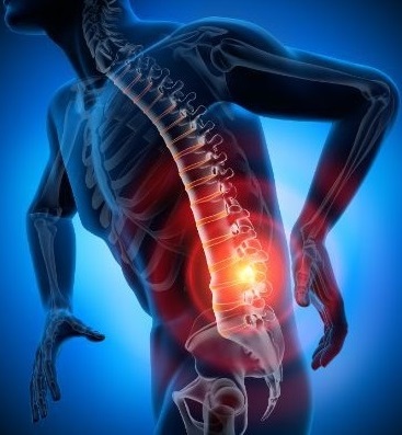
Fractures
Bones are complex structures made up of proteins, minerals and living cells. The skeleton is made up of collagen proteins bound together to form strong, fibrous stems. Minerals are then deposited on this strong foundation. In the case of patients with imperfect osteogenesis, either not enough collagen is produced or what is produced is abnormal.
As a result, the collagen stems are weak and deformed. The bones that contain them break easily, especially in children whose bones are growing rapidly.
Joint Laxity
Laxity is characterized by the instability of the joints that connect two bones. The joints are made up of two bones that are covered by cartilage, tendons and ligaments, which support the bones together and are made up of collagen.
Abnormal collagen present in patients with osteogenesis causes improper functioning of the ligaments, cartilage and tendons, resulting in laxity of the joints.
Blue eye sclera
The eye sclera is a fibrous membrane that covers the eyes and cornea and provides protection. The part of the sclera, which is easy to see, is called the sclera of the eye or the whites of the eye. It contains collagen.
Due to the dysfunction caused by collagen, those diagnosed with fragile type I bone disease have a thinner sclera than normal. This allows the veins in the eye to be seen, which gives the skin a blue color.
Causes of Osteogenesis Imperfecta
A defect in the structure of collagen molecules causes a mutation in the DNA of genes that create collagen. The mutation produces deformed collagen and abnormal collagen fibers. Bones will not be solid because abnormal collagen fibers affect and consume any healthy collagen fiber.
The process is cyclical in a growing child and prevents the formation of new and healthy bones. The result is an increased risk of fractures and ruptures during youth. However, this process will improve towards puberty, when the process of bone growth slows down.
Genetics
Specialists believe that most types of imperfect osteogenesis result from an inherited dominant autosomal pattern. This means that if a child is born to a mother with a dominant mutation, it is 50% likely to be transmitted to the baby. However, it appears that about 15% of children inherited imperfect osteogenesis as a result of the recessive mutation.
In essence, the recessive mutation occurs when both parents are carriers of the recessive gene of the disease, but do not manifest the disorder. The probability of being the carrier of the recessive gene and passing it on to the heirs is 100%, but this does not mean that the disorder will manifest in future generations.
Other factors
An important cause of fragile bone disease is osteoblasts affected by bad collagen molecules. Ossetoblasts are the cells responsible for bone formation. Due to the poor quality of collagen fibers that negatively impact osteoblasts, new bone cells cannot form and divide properly.
Treatment for Osteogenesis Imperfecta
There is currently no known treatment for imperfect osteogenesis. The main goal of treatment should be to prevent accidents and maintain healthy bones.
Children with imperfect osteogenesis should be provided with a high-nutrient diet. They need to exercise regularly and maintain a healthy weight. Often children with imperfect osteogenesis will need splints, a wheelchair, or outpatient care.
Surgical treatment of imperfect osteogenesis depends on the severity of the condition and the patient’s age. In very young children, most fractures are treated as if the child has no underlying condition. However, surgical stabilization of fractures is quite common, even at an early age.
Fractures of children with imperfect osteogenesis are often treated with surgery to stabilize the bones and prevent deformities. One of the procedures used is to insert a metal rod into the center of the hollow bone, to support the fragile bones and to prevent bone deformation.