Fonduri de investiții – 4 aspecte generale pe care trebuie să le cunoști

Jade therapy for back pain

Jade therapy is an extraordinary solution even for those who suffer from advanced disc herniations when the pain goes down on the leg as well as for those in sciatica or lumbago crisis. It is the most performing system globally and offers a profound therapeutic experience. From a therapeutic point of view, it combines a very wide range of principles:

  • Acupressure
  • Thermotherapy
  • Mechanical elongation of the spine
  • Massage

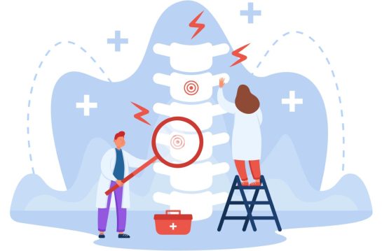
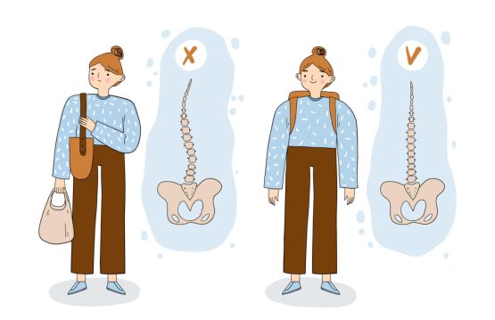
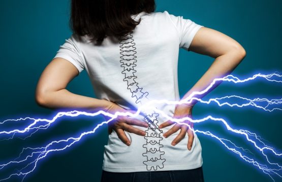
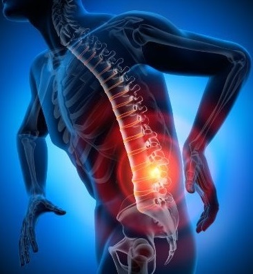
Back pain is pain in any region of the back. It is divided into cervical, thoracic back pain, lower back pain (lumbar) or sacral pain. Additionally, the lumbar region is the most prevalent area for pain as it primarily supports the weight of the upper body.

Episodes of back pain can be acute, sub-acute, or chronic, depending on the duration. Moreover, back pain is one of the most common reasons why people go to the doctor and one of the leading causes of disability worldwide. Most people have back pain at least once in their lifetime.

Jade bed therapy does not require any effort on the part of the patients. A session with this device lasts 45 minutes. Furthermore, a package of 10-12 sessions is recommended for long-term results. Additionally, it is mandatory to consume 250-400 ml of water before the procedure and 250-400 ml of water immediately after the procedure.

The only contraindication for this device is that there is no metal rod implant in the spine.


CONTENT:

  1. How does jade therapy work for back pain?
  2. How an automated Jade massage bed works
  3. Combining elongation procedures with spine toning procedures
  4. Benefits of Jade Therapy
  5. Warning


How does jade therapy work for back pain?

Jade therapy for back pain is based on the principles of heat and pressure therapy. Additionally, this can be done by various methods:

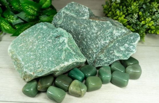
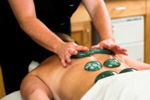
  • Jade stone massage: The therapist may use smooth, pre-heated jade stones to massage painful areas of the back. Furthermore, the heat from jade stones relaxes tense muscles and can help reduce inflammation and discomfort.
  • Jade wraps or compresses: Jade stones can be heated and placed on the affected area or wrapped in cloth to create compresses applied directly to the back. Additionally, this process can help warm the tissues, relax the muscles, and relieve pain.
  • Jade Therapy Pads: There are devices that use jade therapy pads that can be applied to the concerned back. They are designed to emit heat and relax painful areas.


How an automated Jade massage bed works

The action of the massage bed comes in two directions:

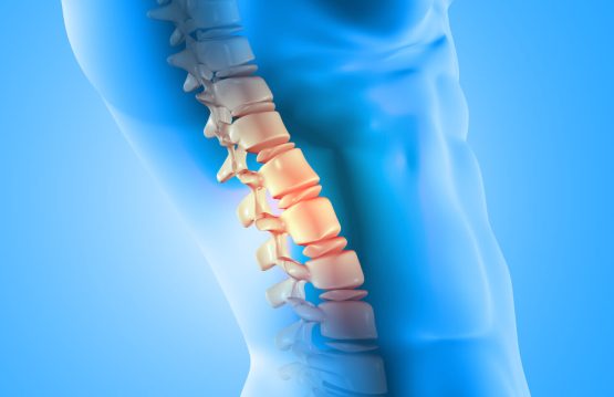
1. Mechanical elongation of the spine as a therapeutic principle

All 7 programs, by reducing the rolls of paravertebral jade, along the spine, according to the natural curvature of the spine, achieve mechanical elongation of the spine with the effect of decompressing the intervertebral discs and increasing the spaces between the vertebrae.

2. Long-term, infrared radiation therapy (Far-Infrared Technology) as a therapeutic principle

Throughout the 7 massage programs, they heat the jade rolls, either based on your personal choice or as per the specialist’s recommendation, to temperatures between 50 and 70 degrees Celsius.

All programs use infrared thermotherapy (heat) and all programs stimulate and improve blood circulation, calming and completely eliminating the symptoms of poor circulation, contributing to a strong detoxification on all 5 pathways, relaxes muscles, eliminates inflammation in the body and contributes strongly to pain therapy, eliminating it.

Combining elongation procedures with spine toning procedures

Through the automated massage bed with Jade therapy, we obtain the elongation of the spine, decompression, and repositioning of the vertebrae. Now, we should ask ourselves why the spine got into the situation where it needed mechanical intervention to eliminate back pain.

In most cases, sedentary lifestyle, overweight and atrophy of the back muscles are the causes that generate these episodes of back pain.

Given that most of you lead a sedentary lifestyle and your daily routine lacks physical activity, heading to the gym may not be the ideal solution for spinal health. Engaging in weightlifting exerts pressure on the spine, potentially causing vertebral compression over time. Furthermore, improperly executed exercises can have lasting adverse effects on the spine.


Benefits of Jade Therapy

  • Deep muscle relaxation
  • Stimulating and improving blood circulation
  • Better oxygenation of muscles, tissues, brain
  • Elimination of insomnia
  • Elimination of emotional problems: depression, panic attacks, anxiety
  • Elimination of BURN OUT syndrome – chronic fatigue, exhaustion, stress
  • Elimination of headaches
  • Elimination of menstrual pain
  • Elimination of back pain
  • Hot spine elongation
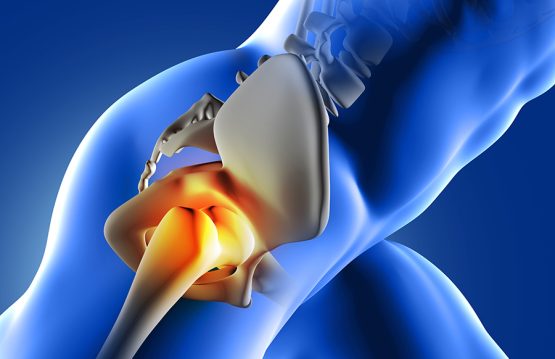
  • Elimination of joint pain
  • Elimination of rheumatic pain
  • Deep detoxification
  • Elimination of chronic constipation
  • Hormonal balance

Warning

Even though jade therapy may help some individuals with back pain, it’s crucial to talk to a doctor or therapist before trying any alternative treatment. Also, make sure it’s safe for your health and avoid using jade stones on irritated or damaged skin.

Diagnosis of back pain

Diagnosis of back pain

In most cases, a specialist is able to make the diagnosis after physical examination and medical history, and imaging tests are done only to confirm the diagnosis and provide additional information.

Low back pain is a common problem, especially in developed countries. These pains can have several causes, so it is recommended to consult a specialist to determine the exact cause of the condition that leads to back pain.

After low back pain, it is advisable to go to the family doctor / general practitioner. He will perform a physical examination and discuss his symptoms and medical history with his patient, and then determine which doctor will refer the patient to.

The diagnosis for a disease presenting symptoms of low back pain can be made by a variety of specialists, including orthopedists, rheumatologists, neurologists, or even internists if there is suspicion that the pain may be related to an organ disease radiating to the lumbar area elsewhere (for example, kidney disease).

A complete medical history and physical examination can usually identify any serious condition that may be causing the pain. During the exam, the doctor will ask about the onset, location, and severity of the pain; duration of symptoms and any limitations in movement; history of previous episodes or any health conditions that may be related to pain.

Along with a detailed examination of the back, neurological tests are performed to determine the cause of the pain and the appropriate treatment. The cause of chronic lower back pain is often difficult to determine even after a thorough examination.

Imaging tests are not justified in most cases. Under certain circumstances, healthcare providers may recommend imaging to rule out specific causes of pain, including tumors and spinal stenosis.


CONTENT:

  1. Magnetic resonance imaging (MRI)
  2. Ultrasound imaging
  3. Bone scans
  4. Blood tests
  5. Radiography
  6. Discography
  7. Computed tomography (CT)
  8. Myelograms


Magnetic resonance imaging (MRI)

It uses a magnetic force instead of radiation to create a computer generated image. Unlike X-rays, which show only bone structures, MRI scans also produce images of soft tissues such as muscles, ligaments, tendons, and blood vessels.

An MRI may be recommended if a problem such as infection, tumor, inflammation, herniated disc, rupture, or nerve compression is suspected. MRI is a non-invasive way to identify a condition that requires prompt surgical treatment. However, in most cases, an MRI scan is not required during the early stages of back pain.


Ultrasound imaging

They are also known as ultrasound scanning or sonography, which utilizes high-frequency sound waves to obtain images inside the body.  Sound wave echoes are recorded and displayed as real-time visual images. Ultrasound imaging can show tears in ligaments, muscles, tendons and other soft tissue masses in the back.


Bone scans

They are used for detecting and monitoring infections, fractures, or bone disorders. A small amount of radioactive material injected into the blood collects in the bones, especially in areas with an abnormality. The images generated by the scanner can identify certain areas of irregular bone metabolism or abnormal blood flow, as well as measure levels of joint disease.


Blood tests

These are not commonly used for diagnosing the cause of back pain. However, in some cases, healthcare providers may perform them to search for indications of inflammation, infection, or the presence of arthritis. Potential tests include erythrocyte sedimentation rate and C-reactive protein.


Radiography

It is often the first imaging technique used to look for broken bones or injured vertebrae. X-rays show bone structure and any vertebral misalignment or fractures. Soft tissues, such as muscles, ligaments, or clogged discs, are not visible in conventional X-rays.


Discography

It can be used when other diagnostic procedures fail to identify the cause of the pain. This procedure involves injecting a contrast dye into a spinal disc suspected to cause low back pain. Fluid pressure on the disc will reproduce the person’s symptoms if that disc is the cause.

The dye will show damaged areas on CT scans performed after injection. Discography can provide useful information in cases where people are considering lumbar surgery or where their pain has not responded to conventional treatments.


Computed tomography (CT)

It visualizes structures of the spine that an X-ray cannot capture, such as a ruptured disc, spinal stenosis, or tumors. Using a computer, CT scanning creates a three-dimensional image from a series of two-dimensional images.


Myelograms

They increase the diagnosis of X-ray and CT images. In this procedure, the doctor injects a contrast dye into the spinal canal to enable visualization of compression of the spinal cord and nerves, caused by herniated discs or fractures, on an X-ray or computerized tomography (CT) scan.

Long-term effects of COVID-19 infection

Long-term effects of COVID-19 infection

COVID-19 infection is proving to be much more than a respiratory illness. It can affect many organs, not just the lungs – from the skin and muscles to the eyes, heart and kidneys – creating long-term health problems. These include fatigue, blurred vision, difficulty breathing, muscle aches, confusion, headaches and even hallucinations.

Most people infected with the new coronavirus tend to have mild to moderate forms of the disease and heal within a few weeks. In contrast, people who survive COVID-19 infection after needing intubation and longer hospitalization may be left with a number of long-term side effects.


CONTENT:

  1. Long-term effects of COVID-19 infection known to date
  2. Long-term side effects – a feature of other viral infections


Long-term effects of COVID-19 infection known to date

Although there is still much to learn from people who have recovered from COVID-19, there are a number of long-term effects that doctors have seen so far.

Lung problems

We know that COVID-19 infection primarily attacks the lungs, causing inflammation. For this reason, survivors may experience long-term breathing difficulties. Some people recovering from COVID-19 may experience a dry cough or pain when breathing, and patients who have needed mechanical ventilation may have more severe symptoms.

Along with the lung pain, there is discomfort in the back area. You shouldn’t be surprised that once you get in touch with the virus, your back starts to hurt.


Neurological problems

Research shows that COVID-19 infection can affect the brain and central nervous system. Some people who have recovered have reported symptoms such as headaches, dizziness, difficulty concentrating or memory, and even hallucinations. Researchers say that these symptoms are more common in patients who have had severe forms of the disease.


Heart problems

A study by Frankford University in Germany showed abnormal heart rates in more than 75% of those studied who recovered from COVID-19. It was found that a considerable majority of patients in the study had inflammation of the heart and mucous membranes. This can be disturbing, as inflammation of the heart can lead to long-term heart disease.

As we are dealing with a relatively new disease, it is not yet known why certain patients experience one or more side effects after infection with the new coronavirus, how long these effects persist and whether COVID-19 infection will cause certain chronic diseases. , who will need lifelong monitoring.


Long-term side effects – a feature of other viral infections

Long-term symptoms of a severe viral illness are not new. Infections with other pathogens are associated with long-lasting symptoms, from heart problems to chronic fatigue. Even common illnesses, such as the flu and pneumonia, can lead to a long recovery. For example, the flu has long been linked to persistent symptoms, such as fatigue and muscle aches.

Infections with certain bacteria and Zika virus, among others, are linked to Guillain-Barre syndrome, a condition in which the immune system attacks the nervous tissue, causing muscle weakness and paralysis.

There is a precedent for infections with other coronaviruses. For example, the first SARS coronavirus and the Middle East Respiratory Virus (MERS) caused serious illness, and a significant number of patients developed acute respiratory disease syndrome (ARDS) and needed intensive care.

Canadian researchers found common issues among SARS survivors in Toronto: sleep disorders, fatigue, depression, and muscle aches. One-third of survivors were forced to change jobs and lifestyles, and only 14% had no long-term symptoms. A Korean study found that 48% of MERS survivors experienced chronic fatigue within a year of recovery.

Many severe COVID-19 patients require a long recovery period, as widely recognized in the medical community. The virus can attack the heart through direct invasion and inflammation, damaging or destroying heart cells and affecting function. Also, the virus disrupts ACE2 receptors, which protect heart cells and degrade angiotensin II, a blood pressure-raising hormone.

Back pain caused by Omicron

Back pain caused by Omicron

Doctors have reported an increase in patients’ back and muscle pain complaints since the onset of the Omicron wave, and data from various countries confirm this.

Since the onset of the coronavirus pandemic, there has been a clear list of symptoms, from fever to difficulty breathing to loss of taste and smell. As the pandemic developed and COVID-19 variants multiplied, the list of symptoms grew.


CONTENT:

  1. Injuries to soft tissues and joints
  2. Why pain occurs
  3. Back pain, the newest symptom of the Omicron variant


Injuries to soft tissues and joints

With COVID-19, the pain complaints of many patients focus on the soft tissues around the joints. Those who have previously had back pain or joint problems may notice an occurrence of these problems after receiving Omicron.

The good news is that for the most part, back and muscle aches decrease and go away on their own when you recover, and the infection disappears from your body, which lasts about two weeks.

Because Omicron is still considered a new strain, there is not enough data to know if back pain will become one of its persistent symptoms. However, if you suffer from persistent back pain that does not improve over time, consult a doctor, whether or not you have had COVID-19.


Why pain occurs

A February report by the World Health Organization (WHO) looked at nearly 56,000 cases of COVID-19 in China and found that almost 15% of patients had muscle pain.
When you get sick, your body triggers an immune response against aggressors, such as harmful bacteria or viruses. Back and leg joint pain is due to the body’s inflammatory response to the virus.

These viral infections cause tremors, chills, body aches and difficulty moving.

This form of back pain feels like cramps or spasms in the back muscles.

Specifically, muscle aches and pains are a result of interleukin-releasing immune cells, which are proteins that help fight invasive pathogens.

Therefore, back pain may be in addition to the usual symptoms of COVID infection, such as fever, dry cough, loss of taste or smell, sore throat, headache or pain in other areas.


Back pain, the newest symptom of the Omicron variant

There are now reports of back pain, a new symptom of the Omicron variant. According to the Jerusalem Post, South Africa, which first reported Omicron, has seen an increase in reports of muscle aches, and especially back pain, among people infected with the virus. Indian and Norwegian data support this claim.

The cause of back pain is probably an increase in the level of inflammation in the body, as the immune system fights the invading virus, which stimulates the muscles and joints. Although it is still too early to know, some doctors suspect that Omicron has a unique effect on the bone and muscle system that is manifested by muscle pain.

While facing a viral infection, the body releases a lot of small proteins called cytokines that activate the immune system against pathogens. Cytokines can stimulate muscles and joints.

However, this virus is not the only one that causes this reaction. Back and muscle pain is a familiar symptom of all sorts of other viral and bacterial infections. This immune response, which is the body’s natural response to a viral infection, can lead to various complaints from patients related to the musculoskeletal system.

Spinal Infections

Spinal Infections

There are spinal infections that can affect the intervertebral discs. When it invades the vertebral plates (the cartilaginous structures that separate the intervertebral disc from the adjacent bone structure) and the vertebral bodies. they are called vertebral osteomyelitis.

If at the time of diagnosis the infection has already compromised 2 structures, it refers to spondylodiscitis. Moreover, this makes the purulent secretion can reach the epidural space (the space around the epidural sac – it is like a sac of tissue, made of dura mater), which causes the spinal cord/its last nerve fibers), causing a spinal epidural abscess.


CONTENT:

  1. What are the causes and factors that predispose to spinal infections?
  2. Symptoms
  3. Evolution and prognosis
  4. Diagnostic
  5. Treatment


What are the causes and factors that predispose to spinal infections?

  • Diabetes
  • Immunocompromised patients (also, organ transplant patients who are being treated with immunosuppressive drugs and intravenous drug users)
  • Alcohol abuse
  • Background surgery on the spine
  • Neoplastic diseases (cancer)
  • Rheumatic diseases
  • Liver cirrhosis
  • Renal insufficiency
  • Minimally invasive non-surgical epidural procedures


Symptoms

At the clinical examination:

  • Additionally, in children, loss of lumbar lordosis, fever, and neurological signs (muscle weakness, paresis, gait disorders) are most common.
  • Adults report local pain in the back – neck / neck, associated with fever and may have neurological deficits – muscle weakness in the legs, urinary incontinence
  • There is a clinical triad for spinal epidural abscess
    1. Back pain
    2. Fever
    3. Neurological deficits (muscle weakness, paresis, sphincter disorders – urinary retention)
  • Paravertebral muscle contracture
  • Root pain (pain that has the path of a nerve)
  • Stiff neck (patient cannot bring chin to chest)


Evolution and prognosis

The unfavorable prognosis in cases of spinal cord infection is associated with:

  • The advanced age of the patient
  • The infection is located in the cervical / thoracic segment of the spine
  • The presence of diabetes
  • Heart disease
  • Neurological disorders (muscle weakness, paresis) that have occurred for more than 36 hours
  • Late diagnosis
  • Infections with bacteria resistant to usual antibiotic treatment


Diagnostic

  • With the help of inflammatory markers analyzed in the blood collected from the patient:
    1. Red blood cell sedimentation rate (ESR)
    2. C-reactive protein level (PCR)
    3. The number of leukocytes
  • Bacteriological cultures of blood and urine collected from the patient
  • Wound culture that has clinical signs of infection
  • Imagistic:
    1. Standard radiography: fast, easy to perform, but has the disadvantage that it does not provide clear evidence in the case of an isolated epidural abscess and does not reveal information about an osteomyelitis in the early stages.
    2. Computed tomography (CT): highlights bone changes caused by an infection, pathological calcifications suggestive of bone tuberculosis.
    3. Contrast-enhanced nuclear magnetic resonance imaging (MRI): Moreover, is the investigation of electives, having a great ability to provide information about the local anatomy, the extent of infection, destruction of adjacent tissues, and possible compression of the spinal cord.


Treatment

NON-SURGICAL TREATMENT

The indication for surgery in patients with spinal infection and neurological impairment (muscle weakness, paresis, sphincter disorders) is not questioned, but there are controversies regarding the choice of treatment for patients with minimal or even without neurological changes.

You can opt for treatment with antibiotics, to which bacteria are sensitive—an aspect established after a previous antibiogram was performed—combined with external immobilization or surgery. Additionally, this depends on the neurological status of the patient at presentation, the severity of the disease, the extent of tissue destruction adjacent, and antibiotic response.

SURGICAL TREATMENT

Surgery is performed immediately if there are signs of compression on the spinal nerves or spinal cord. Moreover, the operation should be considered if there is local pain (back / neck) or in case of changes in the curves of the spine, after non-surgical treatment.

The purpose of surgery is:

  • Additionally, aggressive debridement of tissues affected by infection
  • Collection of biological material for bacteriological examinations
  • Decompression of spinal nerves and spinal cord
  • Drainage of purulent collections from neighboring structures of the spine
Back pain caused by bad habits

Back pain caused by bad habits

Back pain is not just the effect of lifting weights, at the gym or in everyday life, or sleeping on the wrong surface.

Because it affects more and more of the population, being the fifth most common cause of hospitalization and the third most common cause of surgery, it is true that in the United States, Prevention has listed several possible causes.

Knowing and being aware of them can help us correct any mistakes, because, as one overseas chiropractor explains, back pain is “rarely a catastrophic event.”


CONTENT:

  1. Be careful what you exercise!
  2. Stay connected to the office and incorrectly in the car
  3. Your mattress is from another decade
  4. You don’t think about your back when you eat
  5. Stay in your heels for a long time
  6. You carry your whole life in your purse
  7. You don’t relax enough


Be careful what you exercise!

A new study shows that 40% of people with back pain become less active, a completely wrong strategy that will delay healing or worsen the situation.

What you should do: First of all, do sports! Secondly, avoid too many push-ups and abs, which only increase the pain. Related to the abdomen, they can be done, but not to the end and slowly.


Stay connected to the office and incorrectly in the car

Did you know that if you sit on your back, the pressure on your spine is 40% higher than if you were standing?

To be honest, the right office position is probably the last thing you think about when you have to urgently hand in a project.

And certainly the stretching breaks don’t seem like the best activity for the 5-10 minutes for which you allow yourself to detach yourself from the papers. But your back will suffer in these conditions, the unused joints losing their lubrication and aging prematurely.

What you should do: Sit back at 135 degrees to reduce pressure on your spine whenever you talk on the phone or take a minute to relax after making sure your office chair allows you to do that. Keep your head straight while doing this exercise.

Every 30 minutes, get up and take a few steps to get your water, printer papers, or anything that might lift you off your chair.

When driving, be sure to sit at a 90-degree angle to easily reach the pedals. If you sit on your back too much, you may have to stretch your legs too far to reach the pedals, which will put pressure on your spine again.


Your mattress is from another decade

If you don’t remember the last time you replaced it, then it’s time to go shopping. A good mattress lasts 9-10 years, but even so you should change it every 5-7 years, for the good of your back.

What you should do: When choosing a new mattress, make sure it is neither too soft nor too hard. Too much mattress can increase the pressure on the spine and make the pain worse, say Spanish researchers.

To make sure your back is guarded during sleep, put a pillow under your feet, in your knees, when you sleep on your back, one between your legs, if you sleep on one side, or one under your stomach and hips, if you rest on your stomach.


You don’t think about your back when you eat

Research has shown that eating for a healthy heart, for proper weight and for optimal blood sugar is also suitable to prevent back pain.

A study in Finland showed that people with back pain often have clogged arteries to the spine. A healthy blood flow brings nutrients to the spine, taking away what is not useful. If this does not happen, inflammation can occur.

What you should do: Make sure that these inflammations do not occur. Avoid excess caffeine and processed foods (avoid hydrogenated or partially hydrogenated oils) and eat whole grains, soy, nuts and seeds, protein (chicken, fish), vegetables and fruits.


Stay in your heels for a long time

High heels force you to bend your back, putting your muscles to work even more. Even shoes without heels are not healthy, as they can cause back pain.

What you should do: You can wear heels, but not over long distances. Otherwise, keep your shoes in comfortable shoes. A study conducted at Lehigh University on volunteers complaining of back pain showed that after they were given light, flexible shoes, the condition of 80% of them improved considerably within a year.


You carry your whole life in your purse

Even if they are fashionable, large handbags do not keep the shoulders at the same level, the one who wears the purse is below. As the purse tends to always be on the same shoulder, problems will appear sooner or later.

What you should do: Choose a light bag or purse, taking care that when it is loaded. It does not need to exceed 10% of your body weight. If you have a lot of weight in one day, divide the weight between two bags, which you should carry on both shoulders or constantly change the working shoulder.


You don’t relax enough

What is happening in your head, unfortunately, does not just stay there. Stress can cause back pain. If you are tense, constantly stressed, the pain will not be delayed.

What you should do: Laugh with a friend, read a book, or listen to music. Austrian study: Half with herniated disc improved with daily music vs. just painkillers. And the effect was lasting, once this “treatment” was prolonged.

After 21 days, the group of volunteers who had treated themselves through music felt a 40% relief from the pain.

How to prevent spinal deformities

How to prevent spinal deformities

In order to prevent spinal deformities, correct the vicious position of the body, and ensure the health of the back, the specialists recommend gymnastics programs meant to ensure an impeccable posture. It starts with the preparation of muscle tone and the maintenance of joint mobility.

This is based primarily on awareness of our position in the context of daily activities: on the school bench, on the office chair, while driving, when we walk on the street or when we get home.

The training is done through postural recovery and the practice of light maintenance gymnastics programs, performed daily, preferably in the morning and lasting between 15 and 30 minutes. Gymnastic movements or exercises are meant to combat the vicious attitudes that occur, especially in those of us who perform static and sedentary activities.

  •  In the sitting position, the column will be kept straight, possibly supported by the back of the chair;
  •  Do not sit with your torso rotated or tilted, leaning on one elbow. The shoulders should be at the same level;
  • The head rests on the head restraint and is held upright, not projected forward, not rotated or tilted;
  • The legs rest on a stool, the knees being raised at the hips;
  • The arms will be held in a symmetrical position and relaxed on the armrests, at the back or can be crossed on the chest;
  •  When working at the office, the forearms rest on the table at a right angle to the torso, and the chest does not come into contact with the edge of the table;
  • The resting surface must be strong, without allowing the spine to bend during the hours of sleep;
  • You can sleep on the right side, but do not use a large pillow, but a pillow. The upper leg can be slightly bent, avoiding the squatting position;
  • If we have to stand for a long time, we will do lifts on tops and heels, we will perform small steps and short movements, we will not remain motionless in the same place.


CONTENT:

  1. Correct office position
  2. How to lift a weight
  3. Correct position when driving
  4. Other tips to prevent spinal deformities


Correct office position

  • The back of the chair should support our lumbar area and be tilted at an angle of 105 ° to the seat;
  • The screen should be at eye level or just below eye level;
  • The distance between the eyes and the computer should be 80 cm;
  • The angle between the thighs and the legs should be between 90 ° -110 °;
  • The soles must rest on the floor, in no case hanging;
  • Computer support should be straight;
  • Forearms parallel to the ground;
  • The shoulders should be relaxed and the elbows close to the body;
  • At most one hour you have to get up and exercise for 5 minutes.


How to lift a weight

  • Think about exactly where you want to carry the weight before lifting it (you need to make it as easy as possible to be able to transport the object safely);
  • Evaluate the weight of the object you need to lift (grab the object by a corner and lift it slightly);
  • Stay as close to the object as possible;
  • Tighten the abdominal muscles;
  • Bend slightly, bending your knees slightly, keep your back as straight as possible, even slightly arched in the lumbar area;
  • Grasp the object below and at the same time straighten both your back and knees;
  • Do not force yourself and do not make sudden movements;
  • Lift the object with a constant motion;
  • Wear non-slip shoes.


Correct position when driving

  • The knees and elbows should be bent at all times;
  • Shifting gears should be done with your palm over the shifter, without changing the angle of your wrist. Any extra effort affects the tendons, and this is dangerous in the long run;
  • Don’t forget to take regular breaks;
  • The angle between the thigh and the torso should be 100 ° –110 °;
  • The back is glued to the back of the chair.


Other tips to prevent spinal deformities

  • Stay active by exercising regularly. They are important for having strong bones and muscles;
  • Do not subject your body to abuse – alcohol and smoking are harmful to your health;
  • Maintain a healthy mood by having a positive mindset for as long as possible;
  • Lose weight (where appropriate);
  • Give up high heels;
  • Avoid jerky, sudden and strong movements;
  • Do not lift more than 2-3 kg;
  • Avoid prolonged exposure to cold or air conditioning.
Spinal Pain - What you need to know

Spinal Pain – What you need to know

If you are experiencing spinal pain that does not subside within a few days, then you need to see a rheumatologist. Many ailments with unpleasant long-term repercussions can be hidden behind back pain.


CONTENT:

  1. Notions about the anatomy of the spine
  2. Cervical pain
  3. Chest pain
  4. Low back pain


Notions about the anatomy of the spine

The spine is divided into several regions. Depending on the location of the pain, we can figure out which region is affected and what the possible causes are.

The cervical spine is located at the neck and consists of 7 vertebrae.
The thoracic spine includes the shoulders and the back of the torso. It consists of 12 vertebrae.

The lumbar spine is located in the middle and includes 5 vertebrae. The area of ​​the sacro-coccygeal spine is located in the lower back (the sacral column includes 5 vertebrae, and the coccygeal area 4 or 5 vertebrae).


Cervical pain

Cervical pain is located in the back of the neck and can usually occur as a result of sudden movements, after various traumas or secondary to vicious positions.

If neck pain occurs suddenly, it is acute neck pain, probably caused by sudden movements and trauma secondary to ligament damage, displacement of vertebrae and muscle spasm.

Also, talking on the phone or typing while walking will maintain a normal (flexed) position of the cervical spine. Last but not least, inflammation at this level can be a cause of chronic pain syndrome.


Chest pain

Deaf, chronic or acute pain felt like a stab between the shoulders is most likely due to a pathology related to the thoracic spine. Trauma, degenerative diseases, sudden movements and vicious positions are also responsible for the appearance of pain at this level.

People who are overweight may also have acute or chronic chest pain. Another possible cause of localized pain between the shoulders is a herniated disc with various localizations. Skeletal deformities (scoliosis or kyphosis) are also involved in the occurrence of back pain.


Low back pain

Pain in the lumbar spine, or as it is popularly called, is a common and important cause of presentation to a doctor. Osteoporosis and spinal compression can be the cause of low back pain.

Also, inflammatory diseases can affect both the lumbar spine and the sacroiliac spine leading to inflammatory pain at this level. It is characterized by the appearance of pain after rest (usually at night and in the morning). Then, its relief during the day.

Osteoporosis in children

Osteoporosis in children

Osteoporosis in children or juvenile osteoporosis is a condition characterized by a decrease in the concentration of minerals in the bones, which leads to a decrease in bone density and an increased risk of fractures.

In children, osteoporosis is quite rare, and its occurrence is most often caused by certain medications or lifestyle, poor diet and lack of exercise.

Regarding the time of onset and duration of the disease, there is no rule, these being very variable depending on the underlying cause.

What is important in all cases is that this condition is correctly and early diagnosed to prevent complications as much as possible and to give the child a chance to regain bone mass so that the tip of the bone mass is as close as possible. of normal.


CONTENT:

  1. Causes and risk factors
  2. Symptoms of Osteoporosis in children
  3. Treatment of Osteoporosis in children


Causes and risk factors

The causes of juvenile osteoporosis are divided into primary and secondary causes.

The primary causes of juvenile osteoporosis include:

  • imperfect osteogenesis;
  • osteoporosis-pseudoglioma syndrome;
  • Ehlers-Danlos syndrome;
  • Marfan syndrome;
  • Menkes syndrome;
  • idiopathic juvenile osteoporosis.

Secondary causes of juvenile osteoporosis include:

  • chronic inflammatory diseases, such as idiopathic juvenile arthritis and inflammatory bowel disease;
  • chronic malabsorption or eating disorders (eg anorexia nervosa);
  • other chronic diseases, such as chronic kidney disease and cystic fibrosis;
  • endocrine disorders such as hypogonadism,
  • hyperparathyroidism, hyperthyroidism, Cushing’s syndrome, growth hormone deficiency;
  • drugs such as glucocorticoids, anticonvulsants, methotrexate.

Risk factors are, on the one hand, genetic risk factors, which cannot be prevented, and, on the other hand, lifestyle risk factors, which can be prevented and corrected. Lifestyle risk factors include:

  • poor nutrition (especially low calcium and vitamin D intake);
  • lack of physical activity, especially in cases where the child is immobilized, but also excessive physical activity if it causes disorders of the menstrual cycle;
  • smoking;
  • alcohol consumption;
  • excessive caffeine consumption.


Symptoms of Osteoporosis in children

Symptoms appear when the disease is already advanced and the main symptoms experienced by children with this condition are: pain in the back, hips or legs, persistent lameness, compression of the vertebral bodies and a decrease in height. At the same time, there are deformities of the spine.

Juvenile osteoporosis is a significant problem because it occurs during the first years of a child’s bone development.

Osteoporosis is a condition that is often asymptomatic. However, when signs and symptoms occur, they are:

  • pain (usually of the back or lower limbs);
  • deformities of the spine or anterior thorax;
  • decrease in height;
  • difficulty walking, limited mobility, chronic lameness;
  • the occurrence of fractures in low-intensity trauma.

At the same time, children may have various other signs and symptoms, depending on the primary or secondary cause of osteoporosis.


Treatment of Osteoporosis in children

Hospital treatment is rarely necessary and mainly refers to orthopedic treatment (of fractures, but also of any resulting deformities).

Home treatment includes hygienic-dietary measures and possibly drug treatment. Regardless of the cause of osteoporosis, hygienic dietary measures are essential: calcium and vitamin D, regular physical activity (depending on severity, possibly a recovery plan with targeted exercise), smoking cessation, reduction of caffeine consumption, and alcohol.

Activities that can easily lead to trauma and fractures (eg contact sports) should also be avoided. As a treatment, bisphosphonates may be given in severe cases.

A very important aspect of treatment for secondary osteoporosis is the treatment of the underlying cause (including replacement of the drug responsible for osteoporosis or at least dose reduction).


Coxarthrosis: causes, symptoms and treatment

Coxarthrosis: causes, symptoms and treatment

Coxarthrosis is considered to be one of the most serious diseases affecting the global population and not necessarily because of the pain it is accompanied by, but rather from the perspective of how it affects the patient’s quality of life.

The hip joint is one of the most sought after joints in the body. It consists of the femoral head and the acetabular cavity, and between the two there is a protective layer of cartilaginous tissue that has the role of helping the joint to move evenly and easily.

Over time, however, this tissue can deteriorate and lead to the onset of hip osteoarthritis.

Although this condition is found mainly in middle-aged and older patients, it should be noted that it can also occur at younger ages, under 40 years.

It is important to note that it can be cured in the early stages, but many patients ignore the symptoms and thus are forced to resort to surgery and other more invasive forms of treatment. That is why it is advisable to learn to recognize the symptoms, risk factors and causes that lead to the onset of coxarthrosis.


CONTENT:

  1. Causes
  2. Symptoms
  3. Treatment
  4. Prevention


Causes

One of the most common causes of hip osteoarthritis is genetic predisposition. In most cases it is a certain pathology or previous circumstances (a familiar history of hip osteoarthritis, for example).

Other causes of hip osteoarthritis:

  • old age (usually over 60 years old, but, as I mentioned before, these hip pains can also occur at younger ages);
  • various traumas present at the level of the hip (these traumas generally appear as a result of activities that require the joint, such as sports activities);
  • congenital hip dislocation;
  • extra pounds (obesity increases mechanical stress on the hip);
  • gender of patients (men are less affected because their hips are narrower than women);
  • various diseases of the musculoskeletal system that can lead to joint disorders (diabetes, hyperthyroidism, Paget’s disease of the bones, etc.).


Symptoms

Pain is one of the most obvious signs of hip osteoarthritis. It should be noted, however, that this is not a constant pain, but a pain that appears and disappears when the person in question makes an effort.

Other symptoms of coxarthrosis:

  • registration of a certain stiffness at the level of the hip: there are hip pains that can hinder simple activities such as putting on shoes, bending down to pick up an object)
  • the appearance of limping: as the pressure on the hip increases, the tendency to limping can be observed;
  • an obvious decrease in mobility: the patient begins to notice that he can no longer stretch his leg back;
  • muscle atrophy: this consequence occurs mainly against the background of the weakening of the hip joints, which functions as a domino effect;
  • the ability to walk begins to be visibly impaired;
  • the installation of a feeling of fatigue, especially after the patient makes an effort to move or to undertake a certain activity that involves the hip joints;
  • weather-sensitivity: the patient begins to feel more pronounced joint pain when it is raining.


As mentioned above, these hip pains can be easily treated in the early stages of the disease, but patients most often go to the doctor in the advanced stages of the disease.

It is very important, therefore, that when you notice one or more of the above symptoms, you should consult a specialist.


Treatment

Treatment for coxarthrosis begins with a treatment regimen designed to slow the progression of the condition.

If even after this drug treatment there is hip pain and the disability does not improve, then the doctor may also recommend surgery.

In principle, non-surgical treatment may include, in addition to the medication prescribed by the doctor, the observance of advice such as:

  • avoiding long-term efforts involving the hip joint;
  • following physiotherapy and physiotherapy procedures;
  • an adequate diet (especially in cases where the patient is overweight);
  • use a cane to reduce pressure on the hip;
  • periodic rest.


On the other hand, in the case of advanced hip osteoarthritis, the doctor may recommend a complete replacement of the joint (or prosthesis of the hip, as the procedure is also known).

Such an intervention will help the patient to visibly and considerably improve his quality of life.


Prevention

In order to prevent the appearance of this disease, you can take some simple measures to counteract the favorable factors.

Thus, it is recommended to avoid carrying weights, to wear high-heeled shoes, to sit for a very long time on a chair, in low armchairs or standing.

It is also advisable to have a constant exercise program, avoid walking very long distances and always be careful to keep your weight within normal parameters.